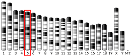
Epiregulin (EPR) is a protein that in humans is encoded by the EREG gene. [5] [6]
Structure
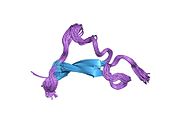
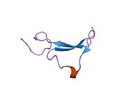
Epiregulin consists of 46 amino acid residues. Its secondary structure contains approximately 30 percent of β-sheet in the strand. [7] Some of the residues form loops and turns due to the hydrogen bonding. [7] The percentage of β-sheet in epiregulin depends on the domain and the secondary structures that they occupy. The polymeric molecules of epiregulin has the formula weight of 5280.1 g/mol with a polypeptide(L), a polymer type. [7]
Structural motifs in most proteins have typical connections in an all β motif. Meaning that the polypeptide chains do not make a crossover connection or in so far as this type of connection has not been observed. Epiregulin is one of the proteins that occupies a typical connection in all β motif. Furthermore, as the structure of epiregulin forms a chain in an all β motif, it also forms β hairpin structural motif. A β hairpin is when the two adjacent anti-parallel β strands connected by a β-turn.
Function
Epiregulin is a member of the epidermal growth factor family. Epiregulin can function as a ligand of epidermal growth factor receptor (EGFR), as well as a ligand of most members of the ERBB (v-erb-b2 oncogene homolog) family of tyrosine-kinase receptors. [6] The secondary structure at the C-terminus epiregulin is different from other epidermal growth factor family ligands because of the lack of hydrogen bonds. The structural difference at the C-terminus may provide an explanation for the reduced binding affinity of epiregulin to the ERBB receptors. [7]
References
- ^ a b c GRCh38: Ensembl release 89: ENSG00000124882 – Ensembl, May 2017
- ^ a b c GRCm38: Ensembl release 89: ENSMUSG00000029377 – Ensembl, May 2017
- ^ "Human PubMed Reference:". National Center for Biotechnology Information, U.S. National Library of Medicine.
- ^ "Mouse PubMed Reference:". National Center for Biotechnology Information, U.S. National Library of Medicine.
- ^ Toyoda H, Komurasaki T, Uchida D, Morimoto S (August 1997). "Distribution of mRNA for human epiregulin, a differentially expressed member of the epidermal growth factor family". Biochem. J. 326 (1): 69–75. doi: 10.1042/bj3260069. PMC 1218638. PMID 9337852.
- ^ a b "Entrez Gene: epiregulin".
- ^ a b c d Sato K, Nakamura T, Mizuguchi M, Miura K, Tada M, Aizawa T, Gomi T, Miyamoto K, Kawano K (October 2003). "Solution structure of epiregulin and the effect of its C-terminal domain for receptor binding affinity". FEBS Lett. 553 (3): 232–8. doi: 10.1016/s0014-5793(03)01005-6. PMID 14572630. S2CID 24761378.
Further reading
- Yamamoto T, Akisue T, Marui T, et al. (2004). "Expression of betacellulin, heparin-binding epidermal growth factor and epiregulin in human malignant fibrous histiocytoma". Anticancer Res. 24 (3b): 2007–10. PMID 15274392.
- Li S, Takeuchi F, Wang JA, et al. (2008). "Mesenchymal-epithelial interactions involving epiregulin in tuberous sclerosis complex hamartomas". Proc. Natl. Acad. Sci. U.S.A. 105 (9): 3539–44. Bibcode: 2008PNAS..105.3539L. doi: 10.1073/pnas.0712397105. PMC 2265180. PMID 18292222.
- Cho MC, Choi HS, Lee S, et al. (2008). "Epiregulin expression by Ets-1 and ERK signaling pathway in Ki-ras-transformed cells". Biochem. Biophys. Res. Commun. 377 (3): 832–7. doi: 10.1016/j.bbrc.2008.10.053. PMID 18948081.
- Lindvall C, Hou M, Komurasaki T, et al. (2003). "Molecular characterization of human telomerase reverse transcriptase-immortalized human fibroblasts by gene expression profiling: activation of the epiregulin gene". Cancer Res. 63 (8): 1743–7. PMID 12702554.
- Morita S, Shirakata Y, Shiraishi A, et al. (2007). "Human corneal epithelial cell proliferation by epiregulin and its cross-induction by other EGF family members". Mol. Vis. 13: 2119–28. PMID 18079685.
- Freimann S, Ben-Ami I, Dantes A, et al. (2004). "EGF-like factor epiregulin and amphiregulin expression is regulated by gonadotropins/cAMP in human ovarian follicular cells". Biochem. Biophys. Res. Commun. 324 (2): 829–34. doi: 10.1016/j.bbrc.2004.09.129. PMID 15474502.
- Ben-Ami I, Armon L, Freimann S, et al. (2009). "EGF-like growth factors as LH mediators in the human corpus luteum". Hum. Reprod. 24 (1): 176–84. doi: 10.1093/humrep/den359. PMID 18835871.
- Shigeishi H, Higashikawa K, Hiraoka M, et al. (2008). "Expression of epiregulin, a novel epidermal growth factor ligand associated with prognosis in human oral squamous cell carcinomas". Oncol. Rep. 19 (6): 1557–64. doi: 10.3892/or.19.6.1557. PMID 18497965.
- Taylor DS, Cheng X, Pawlowski JE, et al. (1999). "Epiregulin is a potent vascular smooth muscle cell-derived mitogen induced by angiotensin II, endothelin-1, and thrombin". Proc. Natl. Acad. Sci. U.S.A. 96 (4): 1633–8. Bibcode: 1999PNAS...96.1633T. doi: 10.1073/pnas.96.4.1633. PMC 15542. PMID 9990076.
- Zhang J, Iwanaga K, Choi KC, et al. (2008). "Intratumoral epiregulin is a marker of advanced disease in non-small cell lung cancer patients and confers invasive properties on EGFR-mutant cells". Cancer Prev Res (Phila). 1 (3): 201–7. doi: 10.1158/1940-6207.CAPR-08-0014. PMC 3375599. PMID 19138957.
- Draper BK, Komurasaki T, Davidson MK, Nanney LB (2003). "Epiregulin is more potent than EGF or TGFalpha in promoting in vitro wound closure due to enhanced ERK/MAPK activation". J. Cell. Biochem. 89 (6): 1126–37. doi: 10.1002/jcb.10584. PMID 12898511. S2CID 24643892.
- Révillion F, Lhotellier V, Hornez L, Bonneterre J, Peyrat JP (January 2008). "ErbB/HER ligands in human breast cancer, and relationships with their receptors, the bio-pathological features and prognosis". Ann. Oncol. 19 (1): 73–80. doi: 10.1093/annonc/mdm431. PMID 17962208.
- Ben-Ami I, Freimann S, Armon L, et al. (2006). "PGE2 up-regulates EGF-like growth factor biosynthesis in human granulosa cells: new insights into the coordination between PGE2 and LH in ovulation". Mol. Hum. Reprod. 12 (10): 593–9. doi: 10.1093/molehr/gal068. PMID 16888076.
- Takahashi M, Hayashi K, Yoshida K, et al. (2003). "Epiregulin as a major autocrine/paracrine factor released from ERK- and p38MAPK-activated vascular smooth muscle cells". Circulation. 108 (20): 2524–9. doi: 10.1161/01.CIR.0000096482.02567.8C. PMID 14581411. S2CID 8012969.
- Lasky-Su J, Neale BM, Franke B, et al. (2008). "Genome-wide association scan of quantitative traits for attention deficit hyperactivity disorder identifies novel associations and confirms candidate gene associations". Am. J. Med. Genet. B Neuropsychiatr. Genet. 147B (8): 1345–54. doi: 10.1002/ajmg.b.30867. PMID 18821565. S2CID 9493672.
- Gupta GP, Nguyen DX, Chiang AC, et al. (2007). "Mediators of vascular remodelling co-opted for sequential steps in lung metastasis". Nature. 446 (7137): 765–70. Bibcode: 2007Natur.446..765G. doi: 10.1038/nature05760. PMID 17429393. S2CID 4420038.
- Shirakata Y, Komurasaki T, Toyoda H, et al. (2000). "Epiregulin, a novel member of the epidermal growth factor family, is an autocrine growth factor in normal human keratinocytes". J. Biol. Chem. 275 (8): 5748–53. doi: 10.1074/jbc.275.8.5748. PMID 10681561.
This article incorporates text from the United States National Library of Medicine, which is in the public domain.
| EREG | |||||||||||||||||||||||||||||||||||||||||||||||||||
|---|---|---|---|---|---|---|---|---|---|---|---|---|---|---|---|---|---|---|---|---|---|---|---|---|---|---|---|---|---|---|---|---|---|---|---|---|---|---|---|---|---|---|---|---|---|---|---|---|---|---|---|
 | |||||||||||||||||||||||||||||||||||||||||||||||||||
| Identifiers | |||||||||||||||||||||||||||||||||||||||||||||||||||
| Aliases | EREG, EPR, ER, Ep, epiregulin | ||||||||||||||||||||||||||||||||||||||||||||||||||
| External IDs | OMIM: 602061; MGI: 107508; HomoloGene: 1097; GeneCards: EREG; OMA: EREG - orthologs | ||||||||||||||||||||||||||||||||||||||||||||||||||
| |||||||||||||||||||||||||||||||||||||||||||||||||||
| |||||||||||||||||||||||||||||||||||||||||||||||||||
| |||||||||||||||||||||||||||||||||||||||||||||||||||
| |||||||||||||||||||||||||||||||||||||||||||||||||||
| |||||||||||||||||||||||||||||||||||||||||||||||||||
| Wikidata | |||||||||||||||||||||||||||||||||||||||||||||||||||
| |||||||||||||||||||||||||||||||||||||||||||||||||||
Epiregulin (EPR) is a protein that in humans is encoded by the EREG gene. [5] [6]
Structure
Epiregulin consists of 46 amino acid residues. Its secondary structure contains approximately 30 percent of β-sheet in the strand. [7] Some of the residues form loops and turns due to the hydrogen bonding. [7] The percentage of β-sheet in epiregulin depends on the domain and the secondary structures that they occupy. The polymeric molecules of epiregulin has the formula weight of 5280.1 g/mol with a polypeptide(L), a polymer type. [7]
Structural motifs in most proteins have typical connections in an all β motif. Meaning that the polypeptide chains do not make a crossover connection or in so far as this type of connection has not been observed. Epiregulin is one of the proteins that occupies a typical connection in all β motif. Furthermore, as the structure of epiregulin forms a chain in an all β motif, it also forms β hairpin structural motif. A β hairpin is when the two adjacent anti-parallel β strands connected by a β-turn.
Function
Epiregulin is a member of the epidermal growth factor family. Epiregulin can function as a ligand of epidermal growth factor receptor (EGFR), as well as a ligand of most members of the ERBB (v-erb-b2 oncogene homolog) family of tyrosine-kinase receptors. [6] The secondary structure at the C-terminus epiregulin is different from other epidermal growth factor family ligands because of the lack of hydrogen bonds. The structural difference at the C-terminus may provide an explanation for the reduced binding affinity of epiregulin to the ERBB receptors. [7]
References
- ^ a b c GRCh38: Ensembl release 89: ENSG00000124882 – Ensembl, May 2017
- ^ a b c GRCm38: Ensembl release 89: ENSMUSG00000029377 – Ensembl, May 2017
- ^ "Human PubMed Reference:". National Center for Biotechnology Information, U.S. National Library of Medicine.
- ^ "Mouse PubMed Reference:". National Center for Biotechnology Information, U.S. National Library of Medicine.
- ^ Toyoda H, Komurasaki T, Uchida D, Morimoto S (August 1997). "Distribution of mRNA for human epiregulin, a differentially expressed member of the epidermal growth factor family". Biochem. J. 326 (1): 69–75. doi: 10.1042/bj3260069. PMC 1218638. PMID 9337852.
- ^ a b "Entrez Gene: epiregulin".
- ^ a b c d Sato K, Nakamura T, Mizuguchi M, Miura K, Tada M, Aizawa T, Gomi T, Miyamoto K, Kawano K (October 2003). "Solution structure of epiregulin and the effect of its C-terminal domain for receptor binding affinity". FEBS Lett. 553 (3): 232–8. doi: 10.1016/s0014-5793(03)01005-6. PMID 14572630. S2CID 24761378.
Further reading
- Yamamoto T, Akisue T, Marui T, et al. (2004). "Expression of betacellulin, heparin-binding epidermal growth factor and epiregulin in human malignant fibrous histiocytoma". Anticancer Res. 24 (3b): 2007–10. PMID 15274392.
- Li S, Takeuchi F, Wang JA, et al. (2008). "Mesenchymal-epithelial interactions involving epiregulin in tuberous sclerosis complex hamartomas". Proc. Natl. Acad. Sci. U.S.A. 105 (9): 3539–44. Bibcode: 2008PNAS..105.3539L. doi: 10.1073/pnas.0712397105. PMC 2265180. PMID 18292222.
- Cho MC, Choi HS, Lee S, et al. (2008). "Epiregulin expression by Ets-1 and ERK signaling pathway in Ki-ras-transformed cells". Biochem. Biophys. Res. Commun. 377 (3): 832–7. doi: 10.1016/j.bbrc.2008.10.053. PMID 18948081.
- Lindvall C, Hou M, Komurasaki T, et al. (2003). "Molecular characterization of human telomerase reverse transcriptase-immortalized human fibroblasts by gene expression profiling: activation of the epiregulin gene". Cancer Res. 63 (8): 1743–7. PMID 12702554.
- Morita S, Shirakata Y, Shiraishi A, et al. (2007). "Human corneal epithelial cell proliferation by epiregulin and its cross-induction by other EGF family members". Mol. Vis. 13: 2119–28. PMID 18079685.
- Freimann S, Ben-Ami I, Dantes A, et al. (2004). "EGF-like factor epiregulin and amphiregulin expression is regulated by gonadotropins/cAMP in human ovarian follicular cells". Biochem. Biophys. Res. Commun. 324 (2): 829–34. doi: 10.1016/j.bbrc.2004.09.129. PMID 15474502.
- Ben-Ami I, Armon L, Freimann S, et al. (2009). "EGF-like growth factors as LH mediators in the human corpus luteum". Hum. Reprod. 24 (1): 176–84. doi: 10.1093/humrep/den359. PMID 18835871.
- Shigeishi H, Higashikawa K, Hiraoka M, et al. (2008). "Expression of epiregulin, a novel epidermal growth factor ligand associated with prognosis in human oral squamous cell carcinomas". Oncol. Rep. 19 (6): 1557–64. doi: 10.3892/or.19.6.1557. PMID 18497965.
- Taylor DS, Cheng X, Pawlowski JE, et al. (1999). "Epiregulin is a potent vascular smooth muscle cell-derived mitogen induced by angiotensin II, endothelin-1, and thrombin". Proc. Natl. Acad. Sci. U.S.A. 96 (4): 1633–8. Bibcode: 1999PNAS...96.1633T. doi: 10.1073/pnas.96.4.1633. PMC 15542. PMID 9990076.
- Zhang J, Iwanaga K, Choi KC, et al. (2008). "Intratumoral epiregulin is a marker of advanced disease in non-small cell lung cancer patients and confers invasive properties on EGFR-mutant cells". Cancer Prev Res (Phila). 1 (3): 201–7. doi: 10.1158/1940-6207.CAPR-08-0014. PMC 3375599. PMID 19138957.
- Draper BK, Komurasaki T, Davidson MK, Nanney LB (2003). "Epiregulin is more potent than EGF or TGFalpha in promoting in vitro wound closure due to enhanced ERK/MAPK activation". J. Cell. Biochem. 89 (6): 1126–37. doi: 10.1002/jcb.10584. PMID 12898511. S2CID 24643892.
- Révillion F, Lhotellier V, Hornez L, Bonneterre J, Peyrat JP (January 2008). "ErbB/HER ligands in human breast cancer, and relationships with their receptors, the bio-pathological features and prognosis". Ann. Oncol. 19 (1): 73–80. doi: 10.1093/annonc/mdm431. PMID 17962208.
- Ben-Ami I, Freimann S, Armon L, et al. (2006). "PGE2 up-regulates EGF-like growth factor biosynthesis in human granulosa cells: new insights into the coordination between PGE2 and LH in ovulation". Mol. Hum. Reprod. 12 (10): 593–9. doi: 10.1093/molehr/gal068. PMID 16888076.
- Takahashi M, Hayashi K, Yoshida K, et al. (2003). "Epiregulin as a major autocrine/paracrine factor released from ERK- and p38MAPK-activated vascular smooth muscle cells". Circulation. 108 (20): 2524–9. doi: 10.1161/01.CIR.0000096482.02567.8C. PMID 14581411. S2CID 8012969.
- Lasky-Su J, Neale BM, Franke B, et al. (2008). "Genome-wide association scan of quantitative traits for attention deficit hyperactivity disorder identifies novel associations and confirms candidate gene associations". Am. J. Med. Genet. B Neuropsychiatr. Genet. 147B (8): 1345–54. doi: 10.1002/ajmg.b.30867. PMID 18821565. S2CID 9493672.
- Gupta GP, Nguyen DX, Chiang AC, et al. (2007). "Mediators of vascular remodelling co-opted for sequential steps in lung metastasis". Nature. 446 (7137): 765–70. Bibcode: 2007Natur.446..765G. doi: 10.1038/nature05760. PMID 17429393. S2CID 4420038.
- Shirakata Y, Komurasaki T, Toyoda H, et al. (2000). "Epiregulin, a novel member of the epidermal growth factor family, is an autocrine growth factor in normal human keratinocytes". J. Biol. Chem. 275 (8): 5748–53. doi: 10.1074/jbc.275.8.5748. PMID 10681561.
This article incorporates text from the United States National Library of Medicine, which is in the public domain.


