| Vpu | |||||||||
|---|---|---|---|---|---|---|---|---|---|
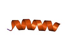
 structure of the channel-forming trans-membrane domain of virus protein "u" (vpu) from
HIV-1 | |||||||||
| Identifiers | |||||||||
| Symbol | Vpu | ||||||||
| Pfam | PF00558 | ||||||||
| InterPro | IPR008187 | ||||||||
| SCOP2 | 1vpu / SCOPe / SUPFAM | ||||||||
| TCDB | 1.A.40 | ||||||||
| OPM superfamily | 262 | ||||||||
| OPM protein | 2k7y | ||||||||
| |||||||||
| Viral Protein Unique | |||||||
|---|---|---|---|---|---|---|---|
| Identifiers | |||||||
| Organism | |||||||
| Symbol | vpu | ||||||
| Entrez | 155945 | ||||||
| RefSeq (Prot) | NP_057855.1 | ||||||
| UniProt | P05919 | ||||||
| Other data | |||||||
| Chromosome | viral genome: 0.01 - 0.01 Mb | ||||||
| |||||||
Vpu is an accessory protein that in HIV is encoded by the vpu gene. Vpu stands for "Viral Protein U". The Vpu protein acts in the degradation of CD4 in the endoplasmic reticulum and in the enhancement of virion release from the plasma membrane of infected cells. [1] Vpu induces the degradation of the CD4 viral receptor and therefore participates in the general downregulation of CD4 expression during the course of HIV infection. Vpu-mediated CD4 degradation is thought to prevent CD4- Env binding in the endoplasmic reticulum to facilitate proper Env assembly into virions. [2] It is found in the membranes of infected cells, but not the virus particles themselves.
The Vpu gene is found exclusively in HIV-1 and some HIV-1-related simian immunodeficiency virus ( SIV) isolates, such as SIVcpz, SIVgsn, and SIVmon, but not in HIV-2 or the majority of SIV isolates. [3] Structural similarities between Vpu and another small viral protein, M2, encoded by influenza A virus were first noted soon after the discovery of Vpu. Since then, Vpu has been shown to form cation-selective ion channels when expressed in Xenopus oocytes or mammalian cells and also when purified and reconstituted into planar lipid bilayers. [4] Vpu also permeabilizes membranes of bacteria and mammalian cells to small molecules. [5] Therefore, it is considered a member of the Viroporins family. [6]
Expression
Vpu and Env are expressed from the same bicistronic mRNA in a Rev-dependent manner, presumably by leaky scanning of ribosomes through the vpu initiation codon. [7] In fact Vpu gene overlaps at its 3′-end with the env gene. Several HIV-1 isolates were found to carry point mutations in the Vpu translation initiation codon but have otherwise intact vpu genes. Since removal of the Vpu initiation codon results in increased expression of the downstream env gene, it is possible that HIV-1 actually uses this mechanism as a molecular switch to regulate the relative expression of Vpu or Env in infected cells. The possible benefits of such a regulation are unclear. [8]
Function
Two main functions have been assigned to the Vpu protein. The first function is known to induce degradation of the viral receptor molecule CD4, and the second function is to enhance the release of newly formed virions from the cell surface. Vpu accomplishes these two functions through two distinct mechanisms. In the case of CD4, Vpu acts as a molecular adaptor to connect CD4 to an E3 ubiquitin ligase complex resulting in CD4 degradation by cellular proteasomes. This requires signals located in Vpu’s cytoplasmic domain. Enhancement of virus release on the other hand involves the neutralization of a cellular host factor, BST-2 (also known as CD317, HM1.24, or tetherin) and requires Vpu’s TM domain. [9] however, the exact mechanism of how Vpu counteracts BST-2 is still unclear. [8] In the absence of Vpu, tetherin binds to the viral envelope and ties it to the cell membrane and other viral particles, impeding release of the viral particles. Recent data suggest that the BST-2 transmembrane domain is crucial for interference by Vpu. The interaction of Vpu and BST-2 results in the downregulation of BST-2 from the cell surface. [10]
BST-2, which is an interferon (IFN)-inducible cell surface protein, appears to “tether” HIV to the cell in the absence of Vpu. BST-2 is a heavily glycosylated 29- to 33-kDa integral membrane protein with both a transmembrane domain and a putative glycosylphosphatidylinositol anchor (GPI). [11] At the cell surface, BST2 resides in lipid rafts through the GPI anchor, whereas its TM domain lies outside them, indirectly interacting with the actin cytoskeleton. Vpu primary site of action is the plasma membrane, where this protein targets cell-surface BST-2 through their mutual TM-to-TM binding, leading to lysosomes, partially dependent on βTrCP. [12]
Structure
Viral protein "u" (Vpu) is an oligomeric, 81-amino acid type I membrane protein (16 kDa) that is translated from vpu-env bicistronic mRNA. The N-terminus of Vpu encoding the transmembrane (TM) anchor represents an active domain important for the regulation of virus release but not CD4 degradation. The C-terminal cytoplasmic domain (54 residues) that contains a pair of serine residues (at positions 52 and 56) constitutively phosphorylated by casein kinase 2. The phosphorylation of two serine residues in the cytoplasmic domain is critical for CD4 degradation in the ER. [13] Based on 2D 1H NMR spectroscopy of a peptide corresponding to the cytoplasmic domain of Vpu, it was proposed that the cytoplasmic domain of Vpu contains two α-helical domains, helix-1 and helix-2, which are connected by an unstructured region containing the two conserved phosphoseryl residues. In addition, computer models predict a third α-helical domain in the transmembrane domain of Vpu, which could play an important role in the formation of ion channels. [14]
See also
References
- ^ Bour S, Schubert U, Strebel K (March 1995). "The human immunodeficiency virus type 1 Vpu protein specifically binds to the cytoplasmic domain of CD4: implications for the mechanism of degradation". Journal of Virology. 69 (3): 1510–20. doi: 10.1128/JVI.69.3.1510-1520.1995. PMC 188742. PMID 7853484.
- ^ Estrabaud E, Le Rouzic E, Lopez-Vergès S, Morel M, Belaïdouni N, Benarous R, Transy C, Berlioz-Torrent C, Margottin-Goguet F (July 2007). "Regulated degradation of the HIV-1 Vpu protein through a betaTrCP-independent pathway limits the release of viral particles". PLOS Pathog. 3 (7): e104. doi: 10.1371/journal.ppat.0030104. PMC 1933454. PMID 17676996.
- ^ Hussain A, Wesley C, Khalid M, Chaudhry A, Jameel S (January 2008). "Human immunodeficiency virus type 1 Vpu protein interacts with CD74 and modulates major histocompatibility complex class II presentation" (PDF). Journal of Virology. 82 (2): 893–902. doi: 10.1128/JVI.01373-07. PMC 2224584. PMID 17959659.
- ^ Ewart GD, Sutherland T, Gage PW, Cox GB (October 1996). "The Vpu protein of human immunodeficiency virus type 1 forms cation-selective ion channels". Journal of Virology. 70 (10): 7108–15. doi: 10.1128/JVI.70.10.7108-7115.1996. PMC 190763. PMID 8794357.
- ^ Gonzalez ME, Carrasco L (September 1998). "The human immunodeficiency virus type 1 Vpu protein enhances membrane permeability". Biochemistry. 37 (39): 13710–9. doi: 10.1021/bi981527f. hdl: 20.500.12105/7976. PMID 9753459.
- ^ Gonzalez ME, Carrasco L (Sep 2003). "Viroporins". FEBS Letters. 552 (1): 28–34. doi: 10.1016/S0014-5793(03)00780-4. hdl: 20.500.12105/7778. PMID 12972148. S2CID 209557930.
- ^ Göttlinger HG, Dorfman T, Cohen EA, Haseltine WA (August 1993). "Vpu protein of human immunodeficiency virus type 1 enhances the release of capsids produced by gag gene constructs of widely divergent retroviruses". Proc. Natl. Acad. Sci. U.S.A. 90 (15): 7381–5. Bibcode: 1993PNAS...90.7381G. doi: 10.1073/pnas.90.15.7381. PMC 47141. PMID 8346259.
- ^ a b Strebel K (December 1996). "Structure and Function of HIV-1 Vpu" (PDF). Los Alamos National Laboratory.
- ^ Andrew A, Strebel K (October 2010). "HIV-1 Vpu targets cell surface markers CD4 and BST-2 through distinct mechanisms". Mol. Aspects Med. 31 (5): 407–17. doi: 10.1016/j.mam.2010.08.002. PMC 2967615. PMID 20858517.
- ^ Andrew AJ, Miyagi E, Strebel K (March 2011). "Differential effects of human immunodeficiency virus type 1 Vpu on the stability of BST-2/tetherin". J. Virol. 85 (6): 2611–9. doi: 10.1128/JVI.02080-10. PMC 3067951. PMID 21191020.
- ^ Douglas JL, Viswanathan K, McCarroll MN, Gustin JK, Früh K, Moses AV (August 2009). "Vpu directs the degradation of the human immunodeficiency virus restriction factor BST-2/Tetherin via a {beta}TrCP-dependent mechanism". J. Virol. 83 (16): 7931–47. doi: 10.1128/JVI.00242-09. PMC 2715753. PMID 19515779.
- ^ Iwabu Y, Fujita H, Kinomoto M, Kaneko K, Ishizaka Y, Tanaka Y, Sata T, Tokunaga K (December 2009). "HIV-1 accessory protein Vpu internalizes cell-surface BST-2/tetherin through transmembrane interactions leading to lysosomes". J. Biol. Chem. 284 (50): 35060–72. doi: 10.1074/jbc.M109.058305. PMC 2787367. PMID 19837671.
- ^ Nomaguchi M, Fujita M, Adachi A (July 2008). "Role of HIV-1 Vpu protein for virus spread and pathogenesis". Microbes Infect. 10 (9): 960–7. doi: 10.1016/j.micinf.2008.07.006. PMID 18672082.
- ^ Cohen EA, Terwilliger EF, Sodroski JG, Haseltine WA (August 1988). "Identification of a protein encoded by the vpu gene of HIV-1". Nature. 334 (6182): 532–4. Bibcode: 1988Natur.334..532C. doi: 10.1038/334532a0. PMID 3043230. S2CID 4372649.
External links
- Genes,+Vpu at the U.S. National Library of Medicine Medical Subject Headings (MeSH)
| Vpu | |||||||||
|---|---|---|---|---|---|---|---|---|---|
 structure of the channel-forming trans-membrane domain of virus protein "u" (vpu) from
HIV-1 | |||||||||
| Identifiers | |||||||||
| Symbol | Vpu | ||||||||
| Pfam | PF00558 | ||||||||
| InterPro | IPR008187 | ||||||||
| SCOP2 | 1vpu / SCOPe / SUPFAM | ||||||||
| TCDB | 1.A.40 | ||||||||
| OPM superfamily | 262 | ||||||||
| OPM protein | 2k7y | ||||||||
| |||||||||
| Viral Protein Unique | |||||||
|---|---|---|---|---|---|---|---|
| Identifiers | |||||||
| Organism | |||||||
| Symbol | vpu | ||||||
| Entrez | 155945 | ||||||
| RefSeq (Prot) | NP_057855.1 | ||||||
| UniProt | P05919 | ||||||
| Other data | |||||||
| Chromosome | viral genome: 0.01 - 0.01 Mb | ||||||
| |||||||
Vpu is an accessory protein that in HIV is encoded by the vpu gene. Vpu stands for "Viral Protein U". The Vpu protein acts in the degradation of CD4 in the endoplasmic reticulum and in the enhancement of virion release from the plasma membrane of infected cells. [1] Vpu induces the degradation of the CD4 viral receptor and therefore participates in the general downregulation of CD4 expression during the course of HIV infection. Vpu-mediated CD4 degradation is thought to prevent CD4- Env binding in the endoplasmic reticulum to facilitate proper Env assembly into virions. [2] It is found in the membranes of infected cells, but not the virus particles themselves.
The Vpu gene is found exclusively in HIV-1 and some HIV-1-related simian immunodeficiency virus ( SIV) isolates, such as SIVcpz, SIVgsn, and SIVmon, but not in HIV-2 or the majority of SIV isolates. [3] Structural similarities between Vpu and another small viral protein, M2, encoded by influenza A virus were first noted soon after the discovery of Vpu. Since then, Vpu has been shown to form cation-selective ion channels when expressed in Xenopus oocytes or mammalian cells and also when purified and reconstituted into planar lipid bilayers. [4] Vpu also permeabilizes membranes of bacteria and mammalian cells to small molecules. [5] Therefore, it is considered a member of the Viroporins family. [6]
Expression
Vpu and Env are expressed from the same bicistronic mRNA in a Rev-dependent manner, presumably by leaky scanning of ribosomes through the vpu initiation codon. [7] In fact Vpu gene overlaps at its 3′-end with the env gene. Several HIV-1 isolates were found to carry point mutations in the Vpu translation initiation codon but have otherwise intact vpu genes. Since removal of the Vpu initiation codon results in increased expression of the downstream env gene, it is possible that HIV-1 actually uses this mechanism as a molecular switch to regulate the relative expression of Vpu or Env in infected cells. The possible benefits of such a regulation are unclear. [8]
Function
Two main functions have been assigned to the Vpu protein. The first function is known to induce degradation of the viral receptor molecule CD4, and the second function is to enhance the release of newly formed virions from the cell surface. Vpu accomplishes these two functions through two distinct mechanisms. In the case of CD4, Vpu acts as a molecular adaptor to connect CD4 to an E3 ubiquitin ligase complex resulting in CD4 degradation by cellular proteasomes. This requires signals located in Vpu’s cytoplasmic domain. Enhancement of virus release on the other hand involves the neutralization of a cellular host factor, BST-2 (also known as CD317, HM1.24, or tetherin) and requires Vpu’s TM domain. [9] however, the exact mechanism of how Vpu counteracts BST-2 is still unclear. [8] In the absence of Vpu, tetherin binds to the viral envelope and ties it to the cell membrane and other viral particles, impeding release of the viral particles. Recent data suggest that the BST-2 transmembrane domain is crucial for interference by Vpu. The interaction of Vpu and BST-2 results in the downregulation of BST-2 from the cell surface. [10]
BST-2, which is an interferon (IFN)-inducible cell surface protein, appears to “tether” HIV to the cell in the absence of Vpu. BST-2 is a heavily glycosylated 29- to 33-kDa integral membrane protein with both a transmembrane domain and a putative glycosylphosphatidylinositol anchor (GPI). [11] At the cell surface, BST2 resides in lipid rafts through the GPI anchor, whereas its TM domain lies outside them, indirectly interacting with the actin cytoskeleton. Vpu primary site of action is the plasma membrane, where this protein targets cell-surface BST-2 through their mutual TM-to-TM binding, leading to lysosomes, partially dependent on βTrCP. [12]
Structure
Viral protein "u" (Vpu) is an oligomeric, 81-amino acid type I membrane protein (16 kDa) that is translated from vpu-env bicistronic mRNA. The N-terminus of Vpu encoding the transmembrane (TM) anchor represents an active domain important for the regulation of virus release but not CD4 degradation. The C-terminal cytoplasmic domain (54 residues) that contains a pair of serine residues (at positions 52 and 56) constitutively phosphorylated by casein kinase 2. The phosphorylation of two serine residues in the cytoplasmic domain is critical for CD4 degradation in the ER. [13] Based on 2D 1H NMR spectroscopy of a peptide corresponding to the cytoplasmic domain of Vpu, it was proposed that the cytoplasmic domain of Vpu contains two α-helical domains, helix-1 and helix-2, which are connected by an unstructured region containing the two conserved phosphoseryl residues. In addition, computer models predict a third α-helical domain in the transmembrane domain of Vpu, which could play an important role in the formation of ion channels. [14]
See also
References
- ^ Bour S, Schubert U, Strebel K (March 1995). "The human immunodeficiency virus type 1 Vpu protein specifically binds to the cytoplasmic domain of CD4: implications for the mechanism of degradation". Journal of Virology. 69 (3): 1510–20. doi: 10.1128/JVI.69.3.1510-1520.1995. PMC 188742. PMID 7853484.
- ^ Estrabaud E, Le Rouzic E, Lopez-Vergès S, Morel M, Belaïdouni N, Benarous R, Transy C, Berlioz-Torrent C, Margottin-Goguet F (July 2007). "Regulated degradation of the HIV-1 Vpu protein through a betaTrCP-independent pathway limits the release of viral particles". PLOS Pathog. 3 (7): e104. doi: 10.1371/journal.ppat.0030104. PMC 1933454. PMID 17676996.
- ^ Hussain A, Wesley C, Khalid M, Chaudhry A, Jameel S (January 2008). "Human immunodeficiency virus type 1 Vpu protein interacts with CD74 and modulates major histocompatibility complex class II presentation" (PDF). Journal of Virology. 82 (2): 893–902. doi: 10.1128/JVI.01373-07. PMC 2224584. PMID 17959659.
- ^ Ewart GD, Sutherland T, Gage PW, Cox GB (October 1996). "The Vpu protein of human immunodeficiency virus type 1 forms cation-selective ion channels". Journal of Virology. 70 (10): 7108–15. doi: 10.1128/JVI.70.10.7108-7115.1996. PMC 190763. PMID 8794357.
- ^ Gonzalez ME, Carrasco L (September 1998). "The human immunodeficiency virus type 1 Vpu protein enhances membrane permeability". Biochemistry. 37 (39): 13710–9. doi: 10.1021/bi981527f. hdl: 20.500.12105/7976. PMID 9753459.
- ^ Gonzalez ME, Carrasco L (Sep 2003). "Viroporins". FEBS Letters. 552 (1): 28–34. doi: 10.1016/S0014-5793(03)00780-4. hdl: 20.500.12105/7778. PMID 12972148. S2CID 209557930.
- ^ Göttlinger HG, Dorfman T, Cohen EA, Haseltine WA (August 1993). "Vpu protein of human immunodeficiency virus type 1 enhances the release of capsids produced by gag gene constructs of widely divergent retroviruses". Proc. Natl. Acad. Sci. U.S.A. 90 (15): 7381–5. Bibcode: 1993PNAS...90.7381G. doi: 10.1073/pnas.90.15.7381. PMC 47141. PMID 8346259.
- ^ a b Strebel K (December 1996). "Structure and Function of HIV-1 Vpu" (PDF). Los Alamos National Laboratory.
- ^ Andrew A, Strebel K (October 2010). "HIV-1 Vpu targets cell surface markers CD4 and BST-2 through distinct mechanisms". Mol. Aspects Med. 31 (5): 407–17. doi: 10.1016/j.mam.2010.08.002. PMC 2967615. PMID 20858517.
- ^ Andrew AJ, Miyagi E, Strebel K (March 2011). "Differential effects of human immunodeficiency virus type 1 Vpu on the stability of BST-2/tetherin". J. Virol. 85 (6): 2611–9. doi: 10.1128/JVI.02080-10. PMC 3067951. PMID 21191020.
- ^ Douglas JL, Viswanathan K, McCarroll MN, Gustin JK, Früh K, Moses AV (August 2009). "Vpu directs the degradation of the human immunodeficiency virus restriction factor BST-2/Tetherin via a {beta}TrCP-dependent mechanism". J. Virol. 83 (16): 7931–47. doi: 10.1128/JVI.00242-09. PMC 2715753. PMID 19515779.
- ^ Iwabu Y, Fujita H, Kinomoto M, Kaneko K, Ishizaka Y, Tanaka Y, Sata T, Tokunaga K (December 2009). "HIV-1 accessory protein Vpu internalizes cell-surface BST-2/tetherin through transmembrane interactions leading to lysosomes". J. Biol. Chem. 284 (50): 35060–72. doi: 10.1074/jbc.M109.058305. PMC 2787367. PMID 19837671.
- ^ Nomaguchi M, Fujita M, Adachi A (July 2008). "Role of HIV-1 Vpu protein for virus spread and pathogenesis". Microbes Infect. 10 (9): 960–7. doi: 10.1016/j.micinf.2008.07.006. PMID 18672082.
- ^ Cohen EA, Terwilliger EF, Sodroski JG, Haseltine WA (August 1988). "Identification of a protein encoded by the vpu gene of HIV-1". Nature. 334 (6182): 532–4. Bibcode: 1988Natur.334..532C. doi: 10.1038/334532a0. PMID 3043230. S2CID 4372649.
External links
- Genes,+Vpu at the U.S. National Library of Medicine Medical Subject Headings (MeSH)