| Capsid protein | |||||||
|---|---|---|---|---|---|---|---|
| Identifiers | |||||||
| Organism | |||||||
| Symbol | C | ||||||
| UniProt | Q9QAB9 | ||||||
| |||||||
| Hepatitis core antigen | |||||||||
|---|---|---|---|---|---|---|---|---|---|
| Identifiers | |||||||||
| Symbol | Hepatitis_core | ||||||||
| Pfam | PF00906 | ||||||||
| InterPro | IPR002006 | ||||||||
| CATH | 7abl | ||||||||
| SCOP2 | 7abl / SCOPe / SUPFAM | ||||||||
| |||||||||


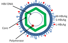
HBcAg (core antigen) is a hepatitis B viral protein. [1] [2] It is an indicator of active viral replication; this means the person infected with Hepatitis B can likely transmit the virus on to another person (i.e. the person is infectious).
Structure and function
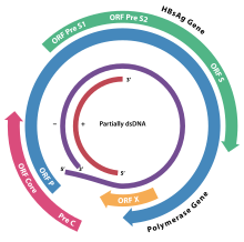
HBcAg is an antigen that can be found on the surface of the nucleocapsid core (the inner most layer of the hepatitis B virus). While both HBcAg and HBeAg are made from the same open reading frame, HBcAg is not secreted. HBcAg is considered "particulate" and it does not circulate in the blood but recent study show it can be detected in serum by Radioimmunoassay. However, it is readily detected in hepatocytes after biopsy. When both HBcAg and HBeAg proteins are present, it acts as a marker of viral replication, and antibodies to these antigens indicates declining replication.
Interactions
Tapasin can interact with HBcAg18-27 and enhance cytotoxic T-lymphocyte response against HBV. [3]
See also
References
- ^ Kimura T, Rokuhara A, Matsumoto A, et al. (May 2003). "New enzyme immunoassay for detection of hepatitis B virus core antigen (HBcAg) and relation between levels of HBcAg and HBV DNA". J. Clin. Microbiol. 41 (5): 1901–6. doi: 10.1128/JCM.41.5.1901-1906.2003. PMC 154683. PMID 12734224.
- ^ Cao T, Meuleman P, Desombere I, Sällberg M, Leroux-Roels G (December 2001). "In vivo inhibition of anti-hepatitis B virus core antigen (HBcAg) immunoglobulin G production by HBcAg-specific CD4(+) Th1-type T-cell clones in a hu-PBL-NOD/SCID mouse model". J. Virol. 75 (23): 11449–56. doi: 10.1128/JVI.75.23.11449-11456.2001. PMC 114731. PMID 11689626.
- ^ Chen X (May 2014). "Tapasin modification on the intracellular epitope HBcAg18-27 enhances HBV-specific CTL immune response and inhibits hepatitis B virus replication in vivo". Lab. Invest. 94 (5): 478–90. doi: 10.1038/labinvest.2014.6. PMID 24614195.
| Capsid protein | |||||||
|---|---|---|---|---|---|---|---|
| Identifiers | |||||||
| Organism | |||||||
| Symbol | C | ||||||
| UniProt | Q9QAB9 | ||||||
| |||||||
| Hepatitis core antigen | |||||||||
|---|---|---|---|---|---|---|---|---|---|
| Identifiers | |||||||||
| Symbol | Hepatitis_core | ||||||||
| Pfam | PF00906 | ||||||||
| InterPro | IPR002006 | ||||||||
| CATH | 7abl | ||||||||
| SCOP2 | 7abl / SCOPe / SUPFAM | ||||||||
| |||||||||


HBcAg (core antigen) is a hepatitis B viral protein. [1] [2] It is an indicator of active viral replication; this means the person infected with Hepatitis B can likely transmit the virus on to another person (i.e. the person is infectious).
Structure and function
HBcAg is an antigen that can be found on the surface of the nucleocapsid core (the inner most layer of the hepatitis B virus). While both HBcAg and HBeAg are made from the same open reading frame, HBcAg is not secreted. HBcAg is considered "particulate" and it does not circulate in the blood but recent study show it can be detected in serum by Radioimmunoassay. However, it is readily detected in hepatocytes after biopsy. When both HBcAg and HBeAg proteins are present, it acts as a marker of viral replication, and antibodies to these antigens indicates declining replication.
Interactions
Tapasin can interact with HBcAg18-27 and enhance cytotoxic T-lymphocyte response against HBV. [3]
See also
References
- ^ Kimura T, Rokuhara A, Matsumoto A, et al. (May 2003). "New enzyme immunoassay for detection of hepatitis B virus core antigen (HBcAg) and relation between levels of HBcAg and HBV DNA". J. Clin. Microbiol. 41 (5): 1901–6. doi: 10.1128/JCM.41.5.1901-1906.2003. PMC 154683. PMID 12734224.
- ^ Cao T, Meuleman P, Desombere I, Sällberg M, Leroux-Roels G (December 2001). "In vivo inhibition of anti-hepatitis B virus core antigen (HBcAg) immunoglobulin G production by HBcAg-specific CD4(+) Th1-type T-cell clones in a hu-PBL-NOD/SCID mouse model". J. Virol. 75 (23): 11449–56. doi: 10.1128/JVI.75.23.11449-11456.2001. PMC 114731. PMID 11689626.
- ^ Chen X (May 2014). "Tapasin modification on the intracellular epitope HBcAg18-27 enhances HBV-specific CTL immune response and inhibits hepatitis B virus replication in vivo". Lab. Invest. 94 (5): 478–90. doi: 10.1038/labinvest.2014.6. PMID 24614195.