| Maxillary vein | |
|---|---|
 Veins of the head and neck. (Internal maxillary vein visible at center.) | |
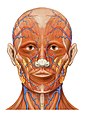
 Lateral head anatomy detail | |
| Details | |
| Drains to | retromandibular vein |
| Artery | maxillary artery |
| Identifiers | |
| Latin | vena maxillaris |
| TA98 | A12.3.05.035 |
| TA2 | 4835 |
| FMA | 70850 |
| Anatomical terminology | |
The maxillary vein or internal maxillary vein is a vein of the head. It is a short trunk which accompanies (the first part of) the maxillary artery. It is formed by a confluence of the veins of the pterygoid plexus. It and passes posterior-ward between the sphenomandibular ligament and the neck of the mandible to enter the parotid gland where unites with the superficial temporal vein to form the retromandibular vein ( posterior facial vein). [1]
Structure
Development
The maxillary vein may be the embryological origin of the central retinal vein. [2]
Additional images
-
 Head anatomy anterior view

References
![]() This article incorporates text in the
public domain from
page 646 of the 20th edition of
Gray's Anatomy (1918)
This article incorporates text in the
public domain from
page 646 of the 20th edition of
Gray's Anatomy (1918)
-
^ Standring, Susan (2020).
Gray's Anatomy: The Anatomical Basis of Clinical Practice (42th ed.). New York. p. 680.
ISBN
978-0-7020-7707-4.
OCLC
1201341621.
{{
cite book}}
: CS1 maint: location missing publisher ( link) - ^ Remington, Lee Ann (2012). "7 - Ocular Embryology". Clinical Anatomy and Physiology of the Visual System (3rd ed.). Butterworth-Heinemann. pp. 123–143. doi: 10.1016/B978-1-4377-1926-0.10007-4. ISBN 978-1-4377-1926-0.
| Maxillary vein | |
|---|---|
|
Veins of the head and neck. (Internal maxillary vein visible at center.) | |
|
Lateral head anatomy detail | |
| Details | |
| Drains to | retromandibular vein |
| Artery | maxillary artery |
| Identifiers | |
| Latin | vena maxillaris |
| TA98 | A12.3.05.035 |
| TA2 | 4835 |
| FMA | 70850 |
| Anatomical terminology | |
The maxillary vein or internal maxillary vein is a vein of the head. It is a short trunk which accompanies (the first part of) the maxillary artery. It is formed by a confluence of the veins of the pterygoid plexus. It and passes posterior-ward between the sphenomandibular ligament and the neck of the mandible to enter the parotid gland where unites with the superficial temporal vein to form the retromandibular vein ( posterior facial vein). [1]
Structure
Development
The maxillary vein may be the embryological origin of the central retinal vein. [2]
Additional images
-
Head anatomy anterior view
References
![]() This article incorporates text in the
public domain from
page 646 of the 20th edition of
Gray's Anatomy (1918)
This article incorporates text in the
public domain from
page 646 of the 20th edition of
Gray's Anatomy (1918)
-
^ Standring, Susan (2020).
Gray's Anatomy: The Anatomical Basis of Clinical Practice (42th ed.). New York. p. 680.
ISBN
978-0-7020-7707-4.
OCLC
1201341621.
{{
cite book}}
: CS1 maint: location missing publisher ( link) - ^ Remington, Lee Ann (2012). "7 - Ocular Embryology". Clinical Anatomy and Physiology of the Visual System (3rd ed.). Butterworth-Heinemann. pp. 123–143. doi: 10.1016/B978-1-4377-1926-0.10007-4. ISBN 978-1-4377-1926-0.