| Thioredoxin reductase | |
|---|---|
| Identifiers | |
| Symbol | ? |
| InterPro | IPR005982 |
| PROSITE | PS00573 |
| SCOP2 | 1zof / SCOPe / SUPFAM |
Thioredoxin reductases (TR, TrxR) ( EC 1.8.1.9) are enzymes that reduce thioredoxin (Trx). [1] Two classes of thioredoxin reductase have been identified: one class in bacteria and some eukaryotes and one in animals. In bacteria TrxR also catalyzes the reduction of glutaredoxin like proteins known as NrdH. [2] [3] [4] Both classes are flavoproteins which function as homodimers. Each monomer contains a FAD prosthetic group, a NADPH binding domain, and an active site containing a redox-active disulfide bond. [5]
Cellular role
Thioredoxin reductases are enzymes that catalyze the reduction of thioredoxin [1] and hence they are a central component in the thioredoxin system. Together with thioredoxin (Trx) and NADPH this system's most general description is as a system for reducing disulfide bonds in cells. Electrons are taken from NADPH via TrxR and are transferred to the active site of Trx, which goes on to reduce protein disulfides or other substrates. [6] The Trx system exists in all living cells and has an evolutionary history tied to DNA as a genetic material, defense against oxidative damage due to oxygen metabolism, and redox signaling using molecules like hydrogen peroxide and nitric oxide. [7] [8]

Diversity
Two classes of thioredoxin reductase have evolved independently:
- A high molecular weight (MW = ~55,000) type containing a selenocysteine residue in its active site has been identified in higher eukaryotes including humans. This TxR is related to glutathione reductase, trypanothione reductase, mercuric reductase and lipoamide dehydrogenase. [5]
- A low molecular weight (MW = ~ 35,000) type has been identified in archaea, bacteria and other eukarya. [5]
These two classes of TrxR have only ~20% sequence identity in the section of primary sequence where they can be reliably aligned. [5] The net reaction of both classes of TrxR is identical but the mechanism of action of each is distinct. [9]
Humans express three thioredoxin reductase isozymes: thioredoxin reductase 1 (TrxR1, cytosolic), thioredoxin reductase 2 (TrxR2, mitochondrial), thioredoxin reductase 3 (TrxR3, testis specific). [10] Each isozyme is encoded by a separate gene:
|
|
| ||||||||||||||||||||||||||||||||||||||||||||||||||||||||||||||||||||||||||||||||||||||||||
Structure
E. coli
In E. coli ThxR there are two binding domains, one for FAD and another for NADPH. The connection between these two domains is a two-stranded anti-parallel β-sheet. [11] Each domain individually is very similar to the analogous domains in glutathione reductase, and lipoamide dehydrogenase but they relative orientation of these domains in ThxR is rotated by 66 degrees. [11] This becomes significant in the enzyme mechanism of action which is described below. ThxR homo-dimerizes with the interface between the two monomers formed by three alpha-helices and two loops. [11] Each monomer can separately bind a molecule of thioredoxin.
-
 Structure of E. coli ThxR dimer bound thioredoxin
 -
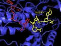
 Structure of E. coli ThxR with FAD and NADPH prosthetic groups labeled


Mammalian
Mammalian TrxR structure is similar to E. coli. It contains a FAD and NADPH binding domain, and an interface between two monomer subunits. In mammalian ThxR there is an insertion in the FAD binding domain between two alpha helices which forms a small pair of beta strands. [12] The active disulfide in the enzyme is located on one of these helices and thus the active disulfide bond is located in the FAD domain and not the NADPH domain as in E. coli and other prokaryotes. [12]
-
 Structure of human ThxR FAD and NADPH prosthetic groups

Mechanism

E. coli
In E. coli ThxR the spatial orientation of the FAD and NADPH domains are such that the redox-active rings of FAD and NADPH are not in close proximity to each other. [1] When the FAD domain of E. coli is rotated 66 degrees with the NADPH domain remaining fixed the two prosthetic groups move into close contact allowing electrons to pass from NADPH to FAD and then to the active site disulfide bond. [1] [15] The conserved active site residues in E. coli are -Cys-Ala-Thr-Cys-. [1]
Mammalian
Mammalian TrxRs have a much higher sequence homology with glutathione reductase than E. coli. [1] The active-site Cys residues in the FAD domain and bound NADPH domain are in close proximity removing the necessity for a 66 degree rotation for electron transfer found in E. coli. An additional feature of the mammalian mechanism is the presence of a selenocysteine residue at the C-terminal end of the protein which is required for catalytic activity. The conserved residues in mammalian active site are -Cys-Val-Asn-Val-Gly-Cys-. [1]
Detection methods
Thioredoxin reductase can be quantified by various methods such as the DTNB assay using Ellman's reagent. The disulfide-based TRFS series of fluorescent probes have shown selective detection of TrxR. [16] [17] [18] [19] Mafireyi synthesized the first diselenide probe that was applied in the detection of TrxR. [20] [21] Other detection methods include immunological techniques and the selenocystine-thioredoxin reductase assay (SC-TR assay).
Clinical significance
Cancer treatment
Since the activity of this enzyme is essential for cell growth and survival, it is a good target for anti-tumor therapy. Furthermore, the enzyme is upregulated in several types of cancer, including malignant mesothelioma. [22] [23] For example, motexafin gadolinium (MGd) is a new chemotherapeutic agent that selectively targets tumor cells, leading to cell death and apoptosis via inhibition of thioredoxin reductase and ribonucleotide reductase.
Cardiomyopathy
Dilated cardiomyopathy ( DCM) is a common diagnosis in cases of congestive heart failure. Thioredoxin reductases are essential proteins for regulating cellular redox balance and mitigating the damage caused by reactive oxygen species generated via oxidative phosphorylation in the mitochondria. Inactivation of mitochondrial TrxR2 in mice results in thinning of the ventricular heart walls and neonatal death. [10] Furthermore two mutations in the TrxR2 gene are found in patients diagnosed with DCM and not in a control population. It is hypothesized that the pathological impact of these mutations is an impaired ability to control oxidative damage in cardiac myocytes. [24]
Antibiotic
There has recently been some research to show that low molecular weight thioredoxin reductase could be a target for novel antibiotics (such as auranofin or Ebselen. [25]) This is especially true for Mycobacterium Haemophilum, and could be used for antibiotic resistant bacteria. [26]
References
- ^ a b c d e f g Mustacich D, Powis G (February 2000). "Thioredoxin reductase". The Biochemical Journal. 346 Pt 1 (1): 1–8. doi: 10.1042/0264-6021:3460001. PMC 1220815. PMID 10657232.
- ^ Jordan A, Aslund F, Pontis E, Reichard P, Holmgren A (July 1997). "Characterization of Escherichia coli NrdH. A glutaredoxin-like protein with a thioredoxin-like activity profile". The Journal of Biological Chemistry. 272 (29): 18044–50. doi: 10.1074/jbc.272.29.18044. PMID 9218434.
- ^ Phulera S, Mande SC (June 2013). "The crystal structure of Mycobacterium tuberculosis NrdH at 0.87 Å suggests a possible mode of its activity". Biochemistry. 52 (23): 4056–65. doi: 10.1021/bi400191z. PMID 23675692.
- ^ Phulera S, Akif M, Sardesai AA, Mande SC (2014-01-01). "Redox Proteins of Mycobacterium tuberculosis". Journal of the Indian Institute of Science. 94 (1): 127–138. ISSN 0970-4140.
- ^ a b c d Hirt RP, Müller S, Embley TM, Coombs GH (July 2002). "The diversity and evolution of thioredoxin reductase: new perspectives". Trends in Parasitology. 18 (7): 302–8. doi: 10.1016/S1471-4922(02)02293-6. PMID 12379950.
- ^ a b Holmgren A, Lu J (May 2010). "Thioredoxin and thioredoxin reductase: current research with special reference to human disease". Biochemical and Biophysical Research Communications. 396 (1): 120–4. doi: 10.1016/j.bbrc.2010.03.083. PMID 20494123.
- ^ Meyer Y, Buchanan BB, Vignols F, Reichheld JP (2009). "Thioredoxins and glutaredoxins: unifying elements in redox biology". Annual Review of Genetics. 43: 335–67. doi: 10.1146/annurev-genet-102108-134201. PMID 19691428.
- ^ Lillig CH, Holmgren A (Jan 2007). "Thioredoxin and related molecules--from biology to health and disease". Antioxidants & Redox Signaling. 9 (1): 25–47. doi: 10.1089/ars.2007.9.25. PMID 17115886.
- ^ Arscott LD, Gromer S, Schirmer RH, Becker K, Williams CH (Apr 1997). "The mechanism of thioredoxin reductase from human placenta is similar to the mechanisms of lipoamide dehydrogenase and glutathione reductase and is distinct from the mechanism of thioredoxin reductase from Escherichia coli". Proceedings of the National Academy of Sciences of the United States of America. 94 (8): 3621–6. Bibcode: 1997PNAS...94.3621A. doi: 10.1073/pnas.94.8.3621. PMC 20490. PMID 9108027.
- ^ a b Conrad M, Jakupoglu C, Moreno SG, Lippl S, Banjac A, Schneider M, Beck H, Hatzopoulos AK, Just U, Sinowatz F, Schmahl W, Chien KR, Wurst W, Bornkamm GW, Brielmeier M (Nov 2004). "Essential role for mitochondrial thioredoxin reductase in hematopoiesis, heart development, and heart function". Molecular and Cellular Biology. 24 (21): 9414–23. doi: 10.1128/MCB.24.21.9414-9423.2004. PMC 522221. PMID 15485910.
- ^ a b c Williams CH (Oct 1995). "Mechanism and structure of thioredoxin reductase from Escherichia coli". FASEB Journal. 9 (13): 1267–76. doi: 10.1096/fasebj.9.13.7557016. hdl: 2027.42/154540. PMID 7557016. S2CID 26055087.
- ^ a b Sandalova T, Zhong L, Lindqvist Y, Holmgren A, Schneider G (Aug 2001). "Three-dimensional structure of a mammalian thioredoxin reductase: implications for mechanism and evolution of a selenocysteine-dependent enzyme". Proceedings of the National Academy of Sciences of the United States of America. 98 (17): 9533–8. Bibcode: 2001PNAS...98.9533S. doi: 10.1073/pnas.171178698. PMC 55487. PMID 11481439.
- ^ Zhong L, Arnér ES, Holmgren A (May 2000). "Structure and mechanism of mammalian thioredoxin reductase: the active site is a redox-active selenolthiol/selenenylsulfide formed from the conserved cysteine-selenocysteine sequence". Proceedings of the National Academy of Sciences of the United States of America. 97 (11): 5854–9. Bibcode: 2000PNAS...97.5854Z. doi: 10.1073/pnas.100114897. PMC 18523. PMID 10801974.
- ^ Becker K, Herold-Mende C, Park JJ, Lowe G, Schirmer RH (Aug 2001). "Human thioredoxin reductase is efficiently inhibited by (2,2':6',2' '-terpyridine)platinum(II) complexes. Possible implications for a novel antitumor strategy". Journal of Medicinal Chemistry. 44 (17): 2784–92. doi: 10.1021/jm001014i. PMID 11495589.
- ^ Lennon BW, Williams CH (Aug 1997). "Reductive half-reaction of thioredoxin reductase from Escherichia coli". Biochemistry. 36 (31): 9464–77. doi: 10.1021/bi970307j. PMID 9235991.
- ^ Li X, Zhang B, Yan C, Li J, Wang S, Wei X, et al. (June 2019). "A fast and specific fluorescent probe for thioredoxin reductase that works via disulphide bond cleavage". Nature Communications. 10 (1): 2745. Bibcode: 2019NatCo..10.2745L. doi: 10.1038/s41467-019-10807-8. PMC 6588570. PMID 31227705.
- ^ Ma H, Zhang J, Zhang Z, Liu Y, Fang J (October 2016). "A fast response and red emission probe for mammalian thioredoxin reductase". Chemical Communications. 52 (81): 12060–12063. doi: 10.1039/C6CC04984B. PMID 27709154.
- ^ Zhao J, Qu Y, Gao H, Zhong M, Li X, Zhang F, et al. (November 2020). "Loss of thioredoxin reductase function in a mouse stroke model disclosed by a two-photon fluorescent probe". Chemical Communications. 56 (90): 14075–14078. doi: 10.1039/D0CC05900E. PMID 33107534. S2CID 225082279.
- ^ Liu Y, Ma H, Zhang L, Cui Y, Liu X, Fang J (February 2016). "A small molecule probe reveals declined mitochondrial thioredoxin reductase activity in a Parkinson's disease model". Chemical Communications. 52 (11): 2296–9. doi: 10.1039/c5cc09998f. PMID 26725656.
- ^ Mafireyi TJ, Laws M, Bassett JW, Cassidy PB, Escobedo JO, Strongin RM (August 2020). "A Diselenide Turn-On Fluorescent Probe for the Detection of Thioredoxin Reductase". Angewandte Chemie. 59 (35): 15147–15151. Bibcode: 2020AngCh.13215259M. doi: 10.1002/ange.202004094. PMC 9438933. PMID 32449244. S2CID 229142596.
- ^ Mafireyi TJ, Escobedo JO, Strongin RM (2021-03-29). "Fluorogenic probes for thioredoxin reductase activity". Results in Chemistry. 3: 100127. doi: 10.1016/j.rechem.2021.100127. ISSN 2211-7156.
- ^ Nilsonne G, Sun X, Nyström C, Rundlöf AK, Potamitou Fernandes A, Björnstedt M, Dobra K (Sep 2006). "Selenite induces apoptosis in sarcomatoid malignant mesothelioma cells through oxidative stress". Free Radical Biology & Medicine. 41 (6): 874–85. doi: 10.1016/j.freeradbiomed.2006.04.031. hdl: 10616/47514. PMID 16934670.
- ^ Kahlos K, Soini Y, Säily M, Koistinen P, Kakko S, Pääkkö P, Holmgren A, Kinnula VL (May 2001). "Up-regulation of thioredoxin and thioredoxin reductase in human malignant pleural mesothelioma". International Journal of Cancer. 95 (3): 198–204. doi: 10.1002/1097-0215(20010520)95:3<198::AID-IJC1034>3.0.CO;2-F. PMID 11307155.
- ^ Sibbing D, Pfeufer A, Perisic T, Mannes AM, Fritz-Wolf K, Unwin S, Sinner MF, Gieger C, Gloeckner CJ, Wichmann HE, Kremmer E, Schäfer Z, Walch A, Hinterseer M, Näbauer M, Kääb S, Kastrati A, Schömig A, Meitinger T, Bornkamm GW, Conrad M, von Beckerath N (May 2011). "Mutations in the mitochondrial thioredoxin reductase gene TXNRD2 cause dilated cardiomyopathy". European Heart Journal. 32 (9): 1121–33. doi: 10.1093/eurheartj/ehq507. hdl: 11858/00-001M-0000-0024-1F10-3. PMID 21247928.
- ^ Marshall AC, Kidd SE, Lamont-Friedrich SJ, Arentz G, Hoffmann P, Coad BR, Bruning JB (March 2019). "Aspergillus fumigatus Thioredoxin Reductase". Antimicrobial Agents and Chemotherapy. 63 (3). doi: 10.1128/AAC.02281-18. PMC 6395915. PMID 30642940.
- ^ Harbut MB, Vilchèze C, Luo X, Hensler ME, Guo H, Yang B, et al. (April 2015). "Auranofin exerts broad-spectrum bactericidal activities by targeting thiol-redox homeostasis". Proceedings of the National Academy of Sciences of the United States of America. 112 (14): 4453–8. Bibcode: 2015PNAS..112.4453H. doi: 10.1073/pnas.1504022112. PMC 4394260. PMID 25831516.
External links
- Thioredoxin+Reductase+(NADPH) at the U.S. National Library of Medicine Medical Subject Headings (MeSH)
| Thioredoxin reductase | |
|---|---|
| Identifiers | |
| Symbol | ? |
| InterPro | IPR005982 |
| PROSITE | PS00573 |
| SCOP2 | 1zof / SCOPe / SUPFAM |
Thioredoxin reductases (TR, TrxR) ( EC 1.8.1.9) are enzymes that reduce thioredoxin (Trx). [1] Two classes of thioredoxin reductase have been identified: one class in bacteria and some eukaryotes and one in animals. In bacteria TrxR also catalyzes the reduction of glutaredoxin like proteins known as NrdH. [2] [3] [4] Both classes are flavoproteins which function as homodimers. Each monomer contains a FAD prosthetic group, a NADPH binding domain, and an active site containing a redox-active disulfide bond. [5]
Cellular role
Thioredoxin reductases are enzymes that catalyze the reduction of thioredoxin [1] and hence they are a central component in the thioredoxin system. Together with thioredoxin (Trx) and NADPH this system's most general description is as a system for reducing disulfide bonds in cells. Electrons are taken from NADPH via TrxR and are transferred to the active site of Trx, which goes on to reduce protein disulfides or other substrates. [6] The Trx system exists in all living cells and has an evolutionary history tied to DNA as a genetic material, defense against oxidative damage due to oxygen metabolism, and redox signaling using molecules like hydrogen peroxide and nitric oxide. [7] [8]
Diversity
Two classes of thioredoxin reductase have evolved independently:
- A high molecular weight (MW = ~55,000) type containing a selenocysteine residue in its active site has been identified in higher eukaryotes including humans. This TxR is related to glutathione reductase, trypanothione reductase, mercuric reductase and lipoamide dehydrogenase. [5]
- A low molecular weight (MW = ~ 35,000) type has been identified in archaea, bacteria and other eukarya. [5]
These two classes of TrxR have only ~20% sequence identity in the section of primary sequence where they can be reliably aligned. [5] The net reaction of both classes of TrxR is identical but the mechanism of action of each is distinct. [9]
Humans express three thioredoxin reductase isozymes: thioredoxin reductase 1 (TrxR1, cytosolic), thioredoxin reductase 2 (TrxR2, mitochondrial), thioredoxin reductase 3 (TrxR3, testis specific). [10] Each isozyme is encoded by a separate gene:
|
|
| ||||||||||||||||||||||||||||||||||||||||||||||||||||||||||||||||||||||||||||||||||||||||||
Structure
E. coli
In E. coli ThxR there are two binding domains, one for FAD and another for NADPH. The connection between these two domains is a two-stranded anti-parallel β-sheet. [11] Each domain individually is very similar to the analogous domains in glutathione reductase, and lipoamide dehydrogenase but they relative orientation of these domains in ThxR is rotated by 66 degrees. [11] This becomes significant in the enzyme mechanism of action which is described below. ThxR homo-dimerizes with the interface between the two monomers formed by three alpha-helices and two loops. [11] Each monomer can separately bind a molecule of thioredoxin.
-
Structure of E. coli ThxR dimer bound thioredoxin
-
Structure of E. coli ThxR with FAD and NADPH prosthetic groups labeled
Mammalian
Mammalian TrxR structure is similar to E. coli. It contains a FAD and NADPH binding domain, and an interface between two monomer subunits. In mammalian ThxR there is an insertion in the FAD binding domain between two alpha helices which forms a small pair of beta strands. [12] The active disulfide in the enzyme is located on one of these helices and thus the active disulfide bond is located in the FAD domain and not the NADPH domain as in E. coli and other prokaryotes. [12]
-
Structure of human ThxR FAD and NADPH prosthetic groups
Mechanism
E. coli
In E. coli ThxR the spatial orientation of the FAD and NADPH domains are such that the redox-active rings of FAD and NADPH are not in close proximity to each other. [1] When the FAD domain of E. coli is rotated 66 degrees with the NADPH domain remaining fixed the two prosthetic groups move into close contact allowing electrons to pass from NADPH to FAD and then to the active site disulfide bond. [1] [15] The conserved active site residues in E. coli are -Cys-Ala-Thr-Cys-. [1]
Mammalian
Mammalian TrxRs have a much higher sequence homology with glutathione reductase than E. coli. [1] The active-site Cys residues in the FAD domain and bound NADPH domain are in close proximity removing the necessity for a 66 degree rotation for electron transfer found in E. coli. An additional feature of the mammalian mechanism is the presence of a selenocysteine residue at the C-terminal end of the protein which is required for catalytic activity. The conserved residues in mammalian active site are -Cys-Val-Asn-Val-Gly-Cys-. [1]
Detection methods
Thioredoxin reductase can be quantified by various methods such as the DTNB assay using Ellman's reagent. The disulfide-based TRFS series of fluorescent probes have shown selective detection of TrxR. [16] [17] [18] [19] Mafireyi synthesized the first diselenide probe that was applied in the detection of TrxR. [20] [21] Other detection methods include immunological techniques and the selenocystine-thioredoxin reductase assay (SC-TR assay).
Clinical significance
Cancer treatment
Since the activity of this enzyme is essential for cell growth and survival, it is a good target for anti-tumor therapy. Furthermore, the enzyme is upregulated in several types of cancer, including malignant mesothelioma. [22] [23] For example, motexafin gadolinium (MGd) is a new chemotherapeutic agent that selectively targets tumor cells, leading to cell death and apoptosis via inhibition of thioredoxin reductase and ribonucleotide reductase.
Cardiomyopathy
Dilated cardiomyopathy ( DCM) is a common diagnosis in cases of congestive heart failure. Thioredoxin reductases are essential proteins for regulating cellular redox balance and mitigating the damage caused by reactive oxygen species generated via oxidative phosphorylation in the mitochondria. Inactivation of mitochondrial TrxR2 in mice results in thinning of the ventricular heart walls and neonatal death. [10] Furthermore two mutations in the TrxR2 gene are found in patients diagnosed with DCM and not in a control population. It is hypothesized that the pathological impact of these mutations is an impaired ability to control oxidative damage in cardiac myocytes. [24]
Antibiotic
There has recently been some research to show that low molecular weight thioredoxin reductase could be a target for novel antibiotics (such as auranofin or Ebselen. [25]) This is especially true for Mycobacterium Haemophilum, and could be used for antibiotic resistant bacteria. [26]
References
- ^ a b c d e f g Mustacich D, Powis G (February 2000). "Thioredoxin reductase". The Biochemical Journal. 346 Pt 1 (1): 1–8. doi: 10.1042/0264-6021:3460001. PMC 1220815. PMID 10657232.
- ^ Jordan A, Aslund F, Pontis E, Reichard P, Holmgren A (July 1997). "Characterization of Escherichia coli NrdH. A glutaredoxin-like protein with a thioredoxin-like activity profile". The Journal of Biological Chemistry. 272 (29): 18044–50. doi: 10.1074/jbc.272.29.18044. PMID 9218434.
- ^ Phulera S, Mande SC (June 2013). "The crystal structure of Mycobacterium tuberculosis NrdH at 0.87 Å suggests a possible mode of its activity". Biochemistry. 52 (23): 4056–65. doi: 10.1021/bi400191z. PMID 23675692.
- ^ Phulera S, Akif M, Sardesai AA, Mande SC (2014-01-01). "Redox Proteins of Mycobacterium tuberculosis". Journal of the Indian Institute of Science. 94 (1): 127–138. ISSN 0970-4140.
- ^ a b c d Hirt RP, Müller S, Embley TM, Coombs GH (July 2002). "The diversity and evolution of thioredoxin reductase: new perspectives". Trends in Parasitology. 18 (7): 302–8. doi: 10.1016/S1471-4922(02)02293-6. PMID 12379950.
- ^ a b Holmgren A, Lu J (May 2010). "Thioredoxin and thioredoxin reductase: current research with special reference to human disease". Biochemical and Biophysical Research Communications. 396 (1): 120–4. doi: 10.1016/j.bbrc.2010.03.083. PMID 20494123.
- ^ Meyer Y, Buchanan BB, Vignols F, Reichheld JP (2009). "Thioredoxins and glutaredoxins: unifying elements in redox biology". Annual Review of Genetics. 43: 335–67. doi: 10.1146/annurev-genet-102108-134201. PMID 19691428.
- ^ Lillig CH, Holmgren A (Jan 2007). "Thioredoxin and related molecules--from biology to health and disease". Antioxidants & Redox Signaling. 9 (1): 25–47. doi: 10.1089/ars.2007.9.25. PMID 17115886.
- ^ Arscott LD, Gromer S, Schirmer RH, Becker K, Williams CH (Apr 1997). "The mechanism of thioredoxin reductase from human placenta is similar to the mechanisms of lipoamide dehydrogenase and glutathione reductase and is distinct from the mechanism of thioredoxin reductase from Escherichia coli". Proceedings of the National Academy of Sciences of the United States of America. 94 (8): 3621–6. Bibcode: 1997PNAS...94.3621A. doi: 10.1073/pnas.94.8.3621. PMC 20490. PMID 9108027.
- ^ a b Conrad M, Jakupoglu C, Moreno SG, Lippl S, Banjac A, Schneider M, Beck H, Hatzopoulos AK, Just U, Sinowatz F, Schmahl W, Chien KR, Wurst W, Bornkamm GW, Brielmeier M (Nov 2004). "Essential role for mitochondrial thioredoxin reductase in hematopoiesis, heart development, and heart function". Molecular and Cellular Biology. 24 (21): 9414–23. doi: 10.1128/MCB.24.21.9414-9423.2004. PMC 522221. PMID 15485910.
- ^ a b c Williams CH (Oct 1995). "Mechanism and structure of thioredoxin reductase from Escherichia coli". FASEB Journal. 9 (13): 1267–76. doi: 10.1096/fasebj.9.13.7557016. hdl: 2027.42/154540. PMID 7557016. S2CID 26055087.
- ^ a b Sandalova T, Zhong L, Lindqvist Y, Holmgren A, Schneider G (Aug 2001). "Three-dimensional structure of a mammalian thioredoxin reductase: implications for mechanism and evolution of a selenocysteine-dependent enzyme". Proceedings of the National Academy of Sciences of the United States of America. 98 (17): 9533–8. Bibcode: 2001PNAS...98.9533S. doi: 10.1073/pnas.171178698. PMC 55487. PMID 11481439.
- ^ Zhong L, Arnér ES, Holmgren A (May 2000). "Structure and mechanism of mammalian thioredoxin reductase: the active site is a redox-active selenolthiol/selenenylsulfide formed from the conserved cysteine-selenocysteine sequence". Proceedings of the National Academy of Sciences of the United States of America. 97 (11): 5854–9. Bibcode: 2000PNAS...97.5854Z. doi: 10.1073/pnas.100114897. PMC 18523. PMID 10801974.
- ^ Becker K, Herold-Mende C, Park JJ, Lowe G, Schirmer RH (Aug 2001). "Human thioredoxin reductase is efficiently inhibited by (2,2':6',2' '-terpyridine)platinum(II) complexes. Possible implications for a novel antitumor strategy". Journal of Medicinal Chemistry. 44 (17): 2784–92. doi: 10.1021/jm001014i. PMID 11495589.
- ^ Lennon BW, Williams CH (Aug 1997). "Reductive half-reaction of thioredoxin reductase from Escherichia coli". Biochemistry. 36 (31): 9464–77. doi: 10.1021/bi970307j. PMID 9235991.
- ^ Li X, Zhang B, Yan C, Li J, Wang S, Wei X, et al. (June 2019). "A fast and specific fluorescent probe for thioredoxin reductase that works via disulphide bond cleavage". Nature Communications. 10 (1): 2745. Bibcode: 2019NatCo..10.2745L. doi: 10.1038/s41467-019-10807-8. PMC 6588570. PMID 31227705.
- ^ Ma H, Zhang J, Zhang Z, Liu Y, Fang J (October 2016). "A fast response and red emission probe for mammalian thioredoxin reductase". Chemical Communications. 52 (81): 12060–12063. doi: 10.1039/C6CC04984B. PMID 27709154.
- ^ Zhao J, Qu Y, Gao H, Zhong M, Li X, Zhang F, et al. (November 2020). "Loss of thioredoxin reductase function in a mouse stroke model disclosed by a two-photon fluorescent probe". Chemical Communications. 56 (90): 14075–14078. doi: 10.1039/D0CC05900E. PMID 33107534. S2CID 225082279.
- ^ Liu Y, Ma H, Zhang L, Cui Y, Liu X, Fang J (February 2016). "A small molecule probe reveals declined mitochondrial thioredoxin reductase activity in a Parkinson's disease model". Chemical Communications. 52 (11): 2296–9. doi: 10.1039/c5cc09998f. PMID 26725656.
- ^ Mafireyi TJ, Laws M, Bassett JW, Cassidy PB, Escobedo JO, Strongin RM (August 2020). "A Diselenide Turn-On Fluorescent Probe for the Detection of Thioredoxin Reductase". Angewandte Chemie. 59 (35): 15147–15151. Bibcode: 2020AngCh.13215259M. doi: 10.1002/ange.202004094. PMC 9438933. PMID 32449244. S2CID 229142596.
- ^ Mafireyi TJ, Escobedo JO, Strongin RM (2021-03-29). "Fluorogenic probes for thioredoxin reductase activity". Results in Chemistry. 3: 100127. doi: 10.1016/j.rechem.2021.100127. ISSN 2211-7156.
- ^ Nilsonne G, Sun X, Nyström C, Rundlöf AK, Potamitou Fernandes A, Björnstedt M, Dobra K (Sep 2006). "Selenite induces apoptosis in sarcomatoid malignant mesothelioma cells through oxidative stress". Free Radical Biology & Medicine. 41 (6): 874–85. doi: 10.1016/j.freeradbiomed.2006.04.031. hdl: 10616/47514. PMID 16934670.
- ^ Kahlos K, Soini Y, Säily M, Koistinen P, Kakko S, Pääkkö P, Holmgren A, Kinnula VL (May 2001). "Up-regulation of thioredoxin and thioredoxin reductase in human malignant pleural mesothelioma". International Journal of Cancer. 95 (3): 198–204. doi: 10.1002/1097-0215(20010520)95:3<198::AID-IJC1034>3.0.CO;2-F. PMID 11307155.
- ^ Sibbing D, Pfeufer A, Perisic T, Mannes AM, Fritz-Wolf K, Unwin S, Sinner MF, Gieger C, Gloeckner CJ, Wichmann HE, Kremmer E, Schäfer Z, Walch A, Hinterseer M, Näbauer M, Kääb S, Kastrati A, Schömig A, Meitinger T, Bornkamm GW, Conrad M, von Beckerath N (May 2011). "Mutations in the mitochondrial thioredoxin reductase gene TXNRD2 cause dilated cardiomyopathy". European Heart Journal. 32 (9): 1121–33. doi: 10.1093/eurheartj/ehq507. hdl: 11858/00-001M-0000-0024-1F10-3. PMID 21247928.
- ^ Marshall AC, Kidd SE, Lamont-Friedrich SJ, Arentz G, Hoffmann P, Coad BR, Bruning JB (March 2019). "Aspergillus fumigatus Thioredoxin Reductase". Antimicrobial Agents and Chemotherapy. 63 (3). doi: 10.1128/AAC.02281-18. PMC 6395915. PMID 30642940.
- ^ Harbut MB, Vilchèze C, Luo X, Hensler ME, Guo H, Yang B, et al. (April 2015). "Auranofin exerts broad-spectrum bactericidal activities by targeting thiol-redox homeostasis". Proceedings of the National Academy of Sciences of the United States of America. 112 (14): 4453–8. Bibcode: 2015PNAS..112.4453H. doi: 10.1073/pnas.1504022112. PMC 4394260. PMID 25831516.
External links
- Thioredoxin+Reductase+(NADPH) at the U.S. National Library of Medicine Medical Subject Headings (MeSH)