|
| This It is of interest to the following WikiProjects: | |||||||||||||||||||||||||||||||||||||||||||||||||||||||||||
| ||||||||||||||||||||||||||||||||||||||||||||||||||||||||||||


.png)

_active.jpg)
In the first image, the curve for L cones is missing the small secondary peak that exists at 410 nm. I'm guessing that the creator of this image simply deleted that part of the curve because he/she thought it would confuse people. However, this secondary peak plays an important role in humans perceiving violet and purple to be similar. — Preceding unsigned comment added by 192.195.76.57 ( talk) 17:48, 24 January 2024 (UTC)
- @ 192.195.76.57 you are either confusing the opsin sensitivities with the color matching functions, which are very different things, or are referring to the beta band of the opsin sensitivity, which only exists in vitro. In vivo, you must incorporate the UV filter of the lens and the beta band disappears, giving only the alpha band, as you see in the image in this article. Curran919 ( talk) 23:14, 24 January 2024 (UTC)
A bit on colour blindness would be interesting. Would it belong here? -- Cuervo 11:34, 18 Apr 2005 (UTC)
Re the comment above, there's a really interesting site at http://www.iamcal.com/toys/colors/index.php (thsi is nothing to do with me) that allows you to select text color and background color and then replicate what folks woudl see if suffering with different forms of color blindness -- I was VERY impressed.
Also, I recently spent a lot of time writing a paper on color vision -- this includes info on cone cells and on the evolution of vision systems.You can find it on my site at www.diycalculator.com/sp-cvision.shtml. I think it provides a very useful background piece that's worth an external link. But I'll leave that up to whoever is in charge. Cheers -- Max (max@diycalculator.com).
and there have been reports of people with four or more types of cone cells This could be the case if the subject is female, since the genes lie on the X chromosome, making it possible, in theory, to have two different kinds of L and M cones, all differing slightly in sensitivity maximum. I do not know if any test exist to test people for this condition, nor is it clear if subjects would benefit and if so to what extent. 88.211.131.57 22:25, 8 February 2007 (UTC) (koenb)
The article states that "Destruction of the cone cells from disease would result in blindness." Should this not read "... would result in colour blindness."? George963 au ( talk) 03:44, 26 July 2017 (UTC)
The article deals chiefly with human cones - would it be reasonable to mention a few things about animal cones? The loss of a cone in the majority of mammals for example, the reclaiming of a third cone by primates through evolution, the fact that many birds in fact possess 4 types of cone, with one having the ability to detect ultraviolet light and giving them a much more complex colour tetrahedron (rather than the triangle of colour available to humans)?
- Agreed. Too bad I know nothing about it. I'd like to see those things added. Twilight Realm 18:49, 9 May 2007 (UTC)
- I just read all about it in The Making of the Fittest. Great story. But long for summarizing here. Dicklyon 05:10, 25 May 2007 (UTC)
Why does "spectral violet" (light with shorter wavelength than blue, as opposed to light with both blue and red wavelengths) appear different than regular blue? There aren't any additional cones to detect sub-blue light. Is it because of the slight increase in sensitivity for red cones for wavelengths shorter than 450 nm? Twilight Realm 18:47, 9 May 2007 (UTC)
When we are seeing 440nm spectral blue, it activates the S cones fully, and also a little bit of of M cones. Our brain is used to treat this as "blue", and it is indeed the color that activates S cones the most. When we see spectral violet, only the S cones are activated, not the M or L ones. That's the difference which makes us see a different color, treated by our brain as "violet", even though it only activates S cones. Ratfox 00:07, 1 November 2007 (UTC)
Indeed, it has been suggested elsewhere that we should think of red, green, and blueviolet cones for this reason. MXVN ( talk) 23:25, 30 August 2012 (UTC)
- Then we'd have to change the name of the red cone to the lime-green cone, since that's typically what 557 nm looks like. We begin to see red when the r/g ratio rises above 5 or so, i.e., red cones stimulated 5 times as much as the green cones. The green cones, on the other hand, are about in the right place for the average young adult with average macular pigment density, which declines with age. (For very young children the purest green is at about 495 nm, for young adults 525, for the elderly about 560. Zyxwv99 ( talk) 00:28, 20 November 2014 (UTC)
Please provide the complete un-normalized response curves of the three types of humans color receptors, and show how this adds up to (or differs from) the Luminosity function.- 69.87.203.133 02:22, 25 May 2007 (UTC)
If there are more rods than cones in the human retina, then rods provide finer details, not cones (as the introduction says). Similarly for the speed, I was taught that rods are faster, again in contrast to the introduction. Pavel —Preceding unsigned comment added by 147.33.113.54 ( talk) 14:57, 6 December 2007 (UTC)
- The fovea is all cones, and that's where you get detail. Cones are fast at high enough light levels; at low light levels where only rods operate, they operate slowly. I'm not sure if what the lead says is really right, though; can you find some sources to check it? Dicklyon ( talk) 15:54, 6 December 2007 (UTC)
Image Cones_SMJ2_E.svg is not found. Link is ok. Is there anyone who can fiy? I don't know how to include images. -- Ernsts ( talk) 15:31, 22 May 2009 (UTC)
It is mentioned in the article (without citation) that S/M/L cones vary from person to person in their specific pigment (and I assume wavelength peak sensitivity). It'd be nice to know more about this -- to what degree do they vary? What impact if any on color perception/discrimination/etc? Might be good to add to the article? Chconnor ( talk) 06:48, 14 September 2014 (UTC)
On the image displaying the cone cell and all of its features, near the top you will see that it says "forms a stacks of regular discs."
Bomb319 ( talk) 04:53, 27 August 2015 (UTC)
Hello fellow Wikipedians,
I have just modified 2 external links on Cone cell. Please take a moment to review my edit. If you have any questions, or need the bot to ignore the links, or the page altogether, please visit this simple FaQ for additional information. I made the following changes:
- Added archive https://web.archive.org/web/20050224231545/http://webvision.umh.es/webvision/photo1.html to http://webvision.umh.es/webvision/photo1.html
- Added archive https://archive.is/20130104021248/http://www.nanobotmodels.com/node/33 to http://www.nanobotmodels.com/node/33
When you have finished reviewing my changes, you may follow the instructions on the template below to fix any issues with the URLs.
This message was posted before February 2018.
After February 2018, "External links modified" talk page sections are no longer generated or monitored by InternetArchiveBot. No special action is required regarding these talk page notices, other than
regular verification using the archive tool instructions below. Editors
have permission to delete these "External links modified" talk page sections if they want to de-clutter talk pages, but see the
RfC before doing mass systematic removals. This message is updated dynamically through the template {{
source check}} (last update: 5 June 2024).
- If you have discovered URLs which were erroneously considered dead by the bot, you can report them with this tool.
- If you found an error with any archives or the URLs themselves, you can fix them with this tool.
Cheers.— InternetArchiveBot ( Report bug) 01:17, 12 August 2017 (UTC)
Cone cells are usually depicted as RGB in colour, according to their sensitivity. But this MUST be wrong. They can only detect absorbed light, not reflected. Are they therefore complementary colours to the light they detect? — Preceding unsigned comment added by Andrewjlockley ( talk • contribs) 21:50, 25 December 2017 (UTC)
- I'm not quite sure what you mean. The cones are not a specific colour, the detect specific colours. Primefac ( talk) 00:13, 2 January 2018 (UTC)
- Yes, they probably do have a bit of color complementary to what they absorb and detect (which are not very close to green and red in the case of the medium- and long-wave-sensitive ones). So the "blue" ones are a bit yellowish or brownish, while the "red" and "green" ones are probably a bit blueish. Or so I guess. Dicklyon ( talk) 01:13, 2 January 2018 (UTC)
The SAME section Structure|Types claims that the peak wavelengths are (about) 560, at 530 & at 420 (nm) and in the same paragraph claims the peaks are 564-580, 534-545 & 420-440!!! Come ON! 40.142.185.236 ( talk) 00:52, 28 February 2018 (UTC)
"Cone cells differ in men and women. The blue cone cell reacts differently in both genders, as a result their perspective of the colour blue is a separate primary colour. The female gender does not have the proper cones required to see the colour blue, they have an entirely different set." First paragraph, with no citations or sources (of course). What in the frick? 2600:1700:6D20:CFF0:4139:7677:3A52:CD20 ( talk) 05:31, 17 February 2020 (UTC)Kerguelen
The article should explain more about the psychophysics, i.e., how (and when) the number and kinds of photoreceptor were learned of, and how the response curves are now determined.
Particularly, Stockman and Sharpe (2000) is an example of recent accepted response curves. It details how the methods have traditionally used detection of a target from a background, and that this method is generally only able to disentangle 2 (not 3) receptor types. Instead, they describe a method of performing genetic sequencing of colorblind participants, to confirm their exact variants of the cone pigments. This allowed them to select participants lacking particular cone types while excluding participants with abnormal remaining cone types. Using these participants they could characterise individual cone types, without having 3 kinds simultaneously interacting. (Those authors also refer to work of Stiles in 1950s, and to other methods that rely on how neural signals from particular cone types are processed differently e.g. using transient stimuli.)
This Neuroscience textbook attributes Thomas Young (scientist) for recognising trichromatic vision, and describes experiments where the participant matches a sample colour by adjusting three superimposed light sources.
Cesiumfrog ( talk) 01:46, 29 October 2020 (UTC)
"The first responds the most to light of longer wavelengths, peaking at about 560 nm; this type is sometimes designated L for long,"
- Any reason we're not calling this the "red cone cell" since, you know, it detects the color red? That's pretty common parlance. 97.122.84.35 ( talk) 00:26, 10 November 2020 (UTC)
I was wondering why red and blue light mixed to violet, so I googled it and found some cone response curves that show the L cone actually being a bit sensitive to spectral violet light as well.
Example: https://commons.m.wikimedia.org/wiki/File:Cone-response-en.svg
But that doesn't match up with the cone response curve in this article. Which one is right, or are they both right somehow? JonathanTybirk ( talk) 13:29, 23 February 2021 (UTC)
- I think your picture would actually be much more appropriate for this article. Keep in mind though that even though the cones themselves are sensitive in the ultraviolet, the lens gradually blocks more and more light as the frequency increases. Where the light gets in practical terms invisible, the ultraviolet starts, conventionally at 400 nm. Aphakic (lenseless) observers however may see well into the ultraviolet, although generally they wear a contact to focus, which blocks ultraviolet. And that may be just as well, because ultraviolet light is bad for the retina; a finch lives only five to ten years or so, but we live a lot longer and would prefer to be able to enjoy the use of our eyes well into old age. — Preceding unsigned comment added by 77.61.180.106 ( talk) 20:59, 7 March 2021 (UTC)
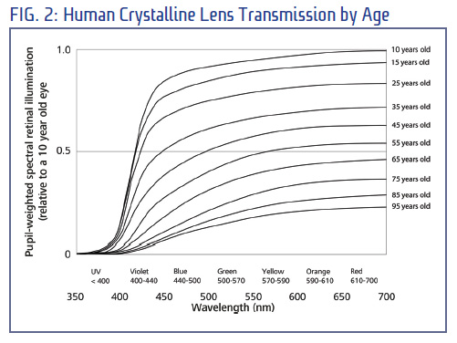
- Bonus: ultraviolet absorbtion by the lens as a function of age: https://2020mag.com/courses/117716/fig2.jpg Note that although one might think colours might appear more and more yellow with age, in practice one gets used to the yellowing and what actually happens is that it becomes harder to distinguish tints, especially in the blues.
- On the red side, the transparent window stretches so far into the infrared that in practice the cone responses themselves are the bottleneck: https://telescope-optics.net/images/retinal_absorption.png — Preceding unsigned comment added by 77.61.180.106 ( talk) 00:13, 8 March 2021 (UTC)
This picture doesn't display the cone response curves, but the S&S2000 LMS curves. Although called ‘cone fundamentals’, they're just a matrix transformation applied to the old CIE XYZ curves, which were based on very crude measurements which were interpolated, extrapolated and standardised with considerable haste. Many of the detail-level features are unphysical and caused by the questionable interpolation method used and the global shape of the curves is probably off as well, especially near the ends of the spectrum. For example, the red and green cones are probably considerably more sensitive in the blue end, with at least the red cones probably having an extra lobe. And if I recall correctly it has recently been discovered that at very long wavelengths, the perceived colour turns from red to slightly orange again, suggesting the green curve is flatter than the red curve there. To be clear, I'm not saying the picture misrepresents the S&S2000 LMS curves, just that this article isn't about that: it's about the cones, so showing different curves is misleading. — Preceding unsigned comment added by 77.61.180.106 ( talk) 20:31, 7 March 2021 (UTC)
- C-Class vital articles
- Wikipedia level-5 vital articles
- Wikipedia vital articles in Biology and health sciences
- C-Class level-5 vital articles
- Wikipedia level-5 vital articles in Biology and health sciences
- C-Class vital articles in Biology and health sciences
- C-Class Physiology articles
- Mid-importance Physiology articles
- Physiology articles about cellular physiology
- WikiProject Physiology articles
- C-Class neuroscience articles
- Mid-importance neuroscience articles
- C-Class Molecular Biology articles
- Unknown-importance Molecular Biology articles
- C-Class MCB articles
- Mid-importance MCB articles
- WikiProject Molecular and Cellular Biology articles
- All WikiProject Molecular Biology pages
- C-Class color articles
- Mid-importance color articles
- All WikiProject Color pages
- C-Class Anatomy articles
- Mid-importance Anatomy articles
- Anatomy articles about microanatomy
- WikiProject Anatomy articles
|
| This It is of interest to the following WikiProjects: | |||||||||||||||||||||||||||||||||||||||||||||||||||||||||||
| ||||||||||||||||||||||||||||||||||||||||||||||||||||||||||||
In the first image, the curve for L cones is missing the small secondary peak that exists at 410 nm. I'm guessing that the creator of this image simply deleted that part of the curve because he/she thought it would confuse people. However, this secondary peak plays an important role in humans perceiving violet and purple to be similar. — Preceding unsigned comment added by 192.195.76.57 ( talk) 17:48, 24 January 2024 (UTC)
- @ 192.195.76.57 you are either confusing the opsin sensitivities with the color matching functions, which are very different things, or are referring to the beta band of the opsin sensitivity, which only exists in vitro. In vivo, you must incorporate the UV filter of the lens and the beta band disappears, giving only the alpha band, as you see in the image in this article. Curran919 ( talk) 23:14, 24 January 2024 (UTC)
A bit on colour blindness would be interesting. Would it belong here? -- Cuervo 11:34, 18 Apr 2005 (UTC)
Re the comment above, there's a really interesting site at http://www.iamcal.com/toys/colors/index.php (thsi is nothing to do with me) that allows you to select text color and background color and then replicate what folks woudl see if suffering with different forms of color blindness -- I was VERY impressed.
Also, I recently spent a lot of time writing a paper on color vision -- this includes info on cone cells and on the evolution of vision systems.You can find it on my site at www.diycalculator.com/sp-cvision.shtml. I think it provides a very useful background piece that's worth an external link. But I'll leave that up to whoever is in charge. Cheers -- Max (max@diycalculator.com).
and there have been reports of people with four or more types of cone cells This could be the case if the subject is female, since the genes lie on the X chromosome, making it possible, in theory, to have two different kinds of L and M cones, all differing slightly in sensitivity maximum. I do not know if any test exist to test people for this condition, nor is it clear if subjects would benefit and if so to what extent. 88.211.131.57 22:25, 8 February 2007 (UTC) (koenb)
The article states that "Destruction of the cone cells from disease would result in blindness." Should this not read "... would result in colour blindness."? George963 au ( talk) 03:44, 26 July 2017 (UTC)
The article deals chiefly with human cones - would it be reasonable to mention a few things about animal cones? The loss of a cone in the majority of mammals for example, the reclaiming of a third cone by primates through evolution, the fact that many birds in fact possess 4 types of cone, with one having the ability to detect ultraviolet light and giving them a much more complex colour tetrahedron (rather than the triangle of colour available to humans)?
- Agreed. Too bad I know nothing about it. I'd like to see those things added. Twilight Realm 18:49, 9 May 2007 (UTC)
- I just read all about it in The Making of the Fittest. Great story. But long for summarizing here. Dicklyon 05:10, 25 May 2007 (UTC)
Why does "spectral violet" (light with shorter wavelength than blue, as opposed to light with both blue and red wavelengths) appear different than regular blue? There aren't any additional cones to detect sub-blue light. Is it because of the slight increase in sensitivity for red cones for wavelengths shorter than 450 nm? Twilight Realm 18:47, 9 May 2007 (UTC)
When we are seeing 440nm spectral blue, it activates the S cones fully, and also a little bit of of M cones. Our brain is used to treat this as "blue", and it is indeed the color that activates S cones the most. When we see spectral violet, only the S cones are activated, not the M or L ones. That's the difference which makes us see a different color, treated by our brain as "violet", even though it only activates S cones. Ratfox 00:07, 1 November 2007 (UTC)
Indeed, it has been suggested elsewhere that we should think of red, green, and blueviolet cones for this reason. MXVN ( talk) 23:25, 30 August 2012 (UTC)
- Then we'd have to change the name of the red cone to the lime-green cone, since that's typically what 557 nm looks like. We begin to see red when the r/g ratio rises above 5 or so, i.e., red cones stimulated 5 times as much as the green cones. The green cones, on the other hand, are about in the right place for the average young adult with average macular pigment density, which declines with age. (For very young children the purest green is at about 495 nm, for young adults 525, for the elderly about 560. Zyxwv99 ( talk) 00:28, 20 November 2014 (UTC)
Please provide the complete un-normalized response curves of the three types of humans color receptors, and show how this adds up to (or differs from) the Luminosity function.- 69.87.203.133 02:22, 25 May 2007 (UTC)
If there are more rods than cones in the human retina, then rods provide finer details, not cones (as the introduction says). Similarly for the speed, I was taught that rods are faster, again in contrast to the introduction. Pavel —Preceding unsigned comment added by 147.33.113.54 ( talk) 14:57, 6 December 2007 (UTC)
- The fovea is all cones, and that's where you get detail. Cones are fast at high enough light levels; at low light levels where only rods operate, they operate slowly. I'm not sure if what the lead says is really right, though; can you find some sources to check it? Dicklyon ( talk) 15:54, 6 December 2007 (UTC)
Image Cones_SMJ2_E.svg is not found. Link is ok. Is there anyone who can fiy? I don't know how to include images. -- Ernsts ( talk) 15:31, 22 May 2009 (UTC)
It is mentioned in the article (without citation) that S/M/L cones vary from person to person in their specific pigment (and I assume wavelength peak sensitivity). It'd be nice to know more about this -- to what degree do they vary? What impact if any on color perception/discrimination/etc? Might be good to add to the article? Chconnor ( talk) 06:48, 14 September 2014 (UTC)
On the image displaying the cone cell and all of its features, near the top you will see that it says "forms a stacks of regular discs."
Bomb319 ( talk) 04:53, 27 August 2015 (UTC)
Hello fellow Wikipedians,
I have just modified 2 external links on Cone cell. Please take a moment to review my edit. If you have any questions, or need the bot to ignore the links, or the page altogether, please visit this simple FaQ for additional information. I made the following changes:
- Added archive https://web.archive.org/web/20050224231545/http://webvision.umh.es/webvision/photo1.html to http://webvision.umh.es/webvision/photo1.html
- Added archive https://archive.is/20130104021248/http://www.nanobotmodels.com/node/33 to http://www.nanobotmodels.com/node/33
When you have finished reviewing my changes, you may follow the instructions on the template below to fix any issues with the URLs.
This message was posted before February 2018.
After February 2018, "External links modified" talk page sections are no longer generated or monitored by InternetArchiveBot. No special action is required regarding these talk page notices, other than
regular verification using the archive tool instructions below. Editors
have permission to delete these "External links modified" talk page sections if they want to de-clutter talk pages, but see the
RfC before doing mass systematic removals. This message is updated dynamically through the template {{
source check}} (last update: 5 June 2024).
- If you have discovered URLs which were erroneously considered dead by the bot, you can report them with this tool.
- If you found an error with any archives or the URLs themselves, you can fix them with this tool.
Cheers.— InternetArchiveBot ( Report bug) 01:17, 12 August 2017 (UTC)
Cone cells are usually depicted as RGB in colour, according to their sensitivity. But this MUST be wrong. They can only detect absorbed light, not reflected. Are they therefore complementary colours to the light they detect? — Preceding unsigned comment added by Andrewjlockley ( talk • contribs) 21:50, 25 December 2017 (UTC)
- I'm not quite sure what you mean. The cones are not a specific colour, the detect specific colours. Primefac ( talk) 00:13, 2 January 2018 (UTC)
- Yes, they probably do have a bit of color complementary to what they absorb and detect (which are not very close to green and red in the case of the medium- and long-wave-sensitive ones). So the "blue" ones are a bit yellowish or brownish, while the "red" and "green" ones are probably a bit blueish. Or so I guess. Dicklyon ( talk) 01:13, 2 January 2018 (UTC)
The SAME section Structure|Types claims that the peak wavelengths are (about) 560, at 530 & at 420 (nm) and in the same paragraph claims the peaks are 564-580, 534-545 & 420-440!!! Come ON! 40.142.185.236 ( talk) 00:52, 28 February 2018 (UTC)
"Cone cells differ in men and women. The blue cone cell reacts differently in both genders, as a result their perspective of the colour blue is a separate primary colour. The female gender does not have the proper cones required to see the colour blue, they have an entirely different set." First paragraph, with no citations or sources (of course). What in the frick? 2600:1700:6D20:CFF0:4139:7677:3A52:CD20 ( talk) 05:31, 17 February 2020 (UTC)Kerguelen
The article should explain more about the psychophysics, i.e., how (and when) the number and kinds of photoreceptor were learned of, and how the response curves are now determined.
Particularly, Stockman and Sharpe (2000) is an example of recent accepted response curves. It details how the methods have traditionally used detection of a target from a background, and that this method is generally only able to disentangle 2 (not 3) receptor types. Instead, they describe a method of performing genetic sequencing of colorblind participants, to confirm their exact variants of the cone pigments. This allowed them to select participants lacking particular cone types while excluding participants with abnormal remaining cone types. Using these participants they could characterise individual cone types, without having 3 kinds simultaneously interacting. (Those authors also refer to work of Stiles in 1950s, and to other methods that rely on how neural signals from particular cone types are processed differently e.g. using transient stimuli.)
This Neuroscience textbook attributes Thomas Young (scientist) for recognising trichromatic vision, and describes experiments where the participant matches a sample colour by adjusting three superimposed light sources.
Cesiumfrog ( talk) 01:46, 29 October 2020 (UTC)
"The first responds the most to light of longer wavelengths, peaking at about 560 nm; this type is sometimes designated L for long,"
- Any reason we're not calling this the "red cone cell" since, you know, it detects the color red? That's pretty common parlance. 97.122.84.35 ( talk) 00:26, 10 November 2020 (UTC)
I was wondering why red and blue light mixed to violet, so I googled it and found some cone response curves that show the L cone actually being a bit sensitive to spectral violet light as well.
Example: https://commons.m.wikimedia.org/wiki/File:Cone-response-en.svg
But that doesn't match up with the cone response curve in this article. Which one is right, or are they both right somehow? JonathanTybirk ( talk) 13:29, 23 February 2021 (UTC)
- I think your picture would actually be much more appropriate for this article. Keep in mind though that even though the cones themselves are sensitive in the ultraviolet, the lens gradually blocks more and more light as the frequency increases. Where the light gets in practical terms invisible, the ultraviolet starts, conventionally at 400 nm. Aphakic (lenseless) observers however may see well into the ultraviolet, although generally they wear a contact to focus, which blocks ultraviolet. And that may be just as well, because ultraviolet light is bad for the retina; a finch lives only five to ten years or so, but we live a lot longer and would prefer to be able to enjoy the use of our eyes well into old age. — Preceding unsigned comment added by 77.61.180.106 ( talk) 20:59, 7 March 2021 (UTC)
- Bonus: ultraviolet absorbtion by the lens as a function of age: https://2020mag.com/courses/117716/fig2.jpg Note that although one might think colours might appear more and more yellow with age, in practice one gets used to the yellowing and what actually happens is that it becomes harder to distinguish tints, especially in the blues.
- On the red side, the transparent window stretches so far into the infrared that in practice the cone responses themselves are the bottleneck: https://telescope-optics.net/images/retinal_absorption.png — Preceding unsigned comment added by 77.61.180.106 ( talk) 00:13, 8 March 2021 (UTC)
This picture doesn't display the cone response curves, but the S&S2000 LMS curves. Although called ‘cone fundamentals’, they're just a matrix transformation applied to the old CIE XYZ curves, which were based on very crude measurements which were interpolated, extrapolated and standardised with considerable haste. Many of the detail-level features are unphysical and caused by the questionable interpolation method used and the global shape of the curves is probably off as well, especially near the ends of the spectrum. For example, the red and green cones are probably considerably more sensitive in the blue end, with at least the red cones probably having an extra lobe. And if I recall correctly it has recently been discovered that at very long wavelengths, the perceived colour turns from red to slightly orange again, suggesting the green curve is flatter than the red curve there. To be clear, I'm not saying the picture misrepresents the S&S2000 LMS curves, just that this article isn't about that: it's about the cones, so showing different curves is misleading. — Preceding unsigned comment added by 77.61.180.106 ( talk) 20:31, 7 March 2021 (UTC)
- C-Class vital articles
- Wikipedia level-5 vital articles
- Wikipedia vital articles in Biology and health sciences
- C-Class level-5 vital articles
- Wikipedia level-5 vital articles in Biology and health sciences
- C-Class vital articles in Biology and health sciences
- C-Class Physiology articles
- Mid-importance Physiology articles
- Physiology articles about cellular physiology
- WikiProject Physiology articles
- C-Class neuroscience articles
- Mid-importance neuroscience articles
- C-Class Molecular Biology articles
- Unknown-importance Molecular Biology articles
- C-Class MCB articles
- Mid-importance MCB articles
- WikiProject Molecular and Cellular Biology articles
- All WikiProject Molecular Biology pages
- C-Class color articles
- Mid-importance color articles
- All WikiProject Color pages
- C-Class Anatomy articles
- Mid-importance Anatomy articles
- Anatomy articles about microanatomy
- WikiProject Anatomy articles