| Muscle fascicle | |
|---|---|
 Structure of a skeletal muscle. (Fascicle labeled at bottom right.) | |
| Details | |
| Part of | Skeletal muscle |
| Identifiers | |
| Latin | fasiculus muscularis |
| TA2 | 2006 |
| TH | H3.03.00.0.00003 |
| Anatomical terminology | |
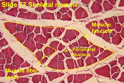
A muscle fascicle is a bundle of skeletal muscle fibers surrounded by perimysium, a type of connective tissue. [1]
Structure
Muscle cells are grouped into muscle fascicles by enveloping perimysium connective tissue. [1] Fascicles are bundled together by epimysium connective tissue. [1] Muscle fascicles typically only contain one type of muscle cell (either type I fibres or type II fibres), but can contain a mixture of both types. [2]
Function
In the heart, specialized cardiac muscle cells transmit electrical impulses from the atrioventricular node (AV node) to the Purkinje fibers – fascicles, also referred to as bundle branches.[ citation needed] These start as a single fascicle of fibers at the AV node called the bundle of His that then splits into three bundle branches: the right fascicular branch, left anterior fascicular branch, and left posterior fascicular branch.
Clinical significance
Myositis may cause thickening of the muscle fascicles. [3] This may be detected with ultrasound scans. [3]
Muscle fascicle structure is a useful diagnostic tool for dermatomyositis. Myocytes towards the edges of the muscle fascicle are typically narrower, while those at the centre of the muscle fascicle are a normal thickness. [4]
Muscle fascicles may be involved in myokymia, although commonly only individual myocytes are involved. [5]
See also
References
- ^ a b c Damjanov, Ivan (2009-01-01), Damjanov, Ivan (ed.), "Chapter 21 - Skeletal Muscles", Pathology Secrets (Third Edition), Philadelphia: Mosby, pp. 434–447, doi: 10.1016/b978-0-323-05594-9.00021-0, ISBN 978-0-323-05594-9, retrieved 2020-11-04
- ^ Gandevia, SIMON C.; Burke, DAVID (2004-01-01), Paxinos, GEORGE; Mai, JÜRGEN K. (eds.), "CHAPTER 5 - Peripheral Motor System", The Human Nervous System (Second Edition), San Diego: Academic Press, pp. 113–133, doi: 10.1016/b978-012547626-3/50006-5, ISBN 978-0-12-547626-3, retrieved 2020-11-04
- ^ a b Möller, Ingrid; Bong, David; Mendieta, Eugenio de Miguel (2010-01-01), Wakefield, Richard J.; D'Agostino, Maria Antonietta (eds.), "Chapter 19 - Soft Tissue Rheumatism", Essential Applications of Musculoskeletal Ultrasound in Rheumatology, Philadelphia: W.B. Saunders, pp. 219–235, doi: 10.1016/b978-1-4377-0127-2.10019-x, ISBN 978-1-4377-0127-2, retrieved 2020-11-04
- ^ Harati, Yadollah; Biliciler, Suur (2010-01-01), Rolak, Loren A. (ed.), "CHAPTER 4 - Myopathies", Neurology Secrets (Fifth Edition), Philadelphia: Mosby, pp. 63–82, doi: 10.1016/b978-0-323-05712-7.00004-0, ISBN 978-0-323-05712-7, retrieved 2020-11-13
- ^ Ha, Ainhi D.; Jankovic, Joseph (2011-01-01), Brotchie, Jonathan; Bezard, Erwan; Jenner, Peter (eds.), "An Introduction to Dyskinesia—The Clinical Spectrum", International Review of Neurobiology, Pathophysiology, Pharmacology, and Biochemistry of Dyskinesia, 98, Academic Press: 1–29, doi: 10.1016/b978-0-12-381328-2.00001-8, ISBN 9780123813282, PMID 21907081, retrieved 2020-11-13
External links
- Histology image: 77_04 at the University of Oklahoma Health Sciences Center – "Slide 77 skeletal muscle"
- Anatomy Atlases – Microscopic Anatomy, plate 05.83 – "Smooth Muscle"
- Diagram at kctcs.edu
|
| This muscle article is a stub. You can help Wikipedia by expanding it. |
| Muscle fascicle | |
|---|---|
|
Structure of a skeletal muscle. (Fascicle labeled at bottom right.) | |
| Details | |
| Part of | Skeletal muscle |
| Identifiers | |
| Latin | fasiculus muscularis |
| TA2 | 2006 |
| TH | H3.03.00.0.00003 |
| Anatomical terminology | |
A muscle fascicle is a bundle of skeletal muscle fibers surrounded by perimysium, a type of connective tissue. [1]
Structure
Muscle cells are grouped into muscle fascicles by enveloping perimysium connective tissue. [1] Fascicles are bundled together by epimysium connective tissue. [1] Muscle fascicles typically only contain one type of muscle cell (either type I fibres or type II fibres), but can contain a mixture of both types. [2]
Function
In the heart, specialized cardiac muscle cells transmit electrical impulses from the atrioventricular node (AV node) to the Purkinje fibers – fascicles, also referred to as bundle branches.[ citation needed] These start as a single fascicle of fibers at the AV node called the bundle of His that then splits into three bundle branches: the right fascicular branch, left anterior fascicular branch, and left posterior fascicular branch.
Clinical significance
Myositis may cause thickening of the muscle fascicles. [3] This may be detected with ultrasound scans. [3]
Muscle fascicle structure is a useful diagnostic tool for dermatomyositis. Myocytes towards the edges of the muscle fascicle are typically narrower, while those at the centre of the muscle fascicle are a normal thickness. [4]
Muscle fascicles may be involved in myokymia, although commonly only individual myocytes are involved. [5]
See also
References
- ^ a b c Damjanov, Ivan (2009-01-01), Damjanov, Ivan (ed.), "Chapter 21 - Skeletal Muscles", Pathology Secrets (Third Edition), Philadelphia: Mosby, pp. 434–447, doi: 10.1016/b978-0-323-05594-9.00021-0, ISBN 978-0-323-05594-9, retrieved 2020-11-04
- ^ Gandevia, SIMON C.; Burke, DAVID (2004-01-01), Paxinos, GEORGE; Mai, JÜRGEN K. (eds.), "CHAPTER 5 - Peripheral Motor System", The Human Nervous System (Second Edition), San Diego: Academic Press, pp. 113–133, doi: 10.1016/b978-012547626-3/50006-5, ISBN 978-0-12-547626-3, retrieved 2020-11-04
- ^ a b Möller, Ingrid; Bong, David; Mendieta, Eugenio de Miguel (2010-01-01), Wakefield, Richard J.; D'Agostino, Maria Antonietta (eds.), "Chapter 19 - Soft Tissue Rheumatism", Essential Applications of Musculoskeletal Ultrasound in Rheumatology, Philadelphia: W.B. Saunders, pp. 219–235, doi: 10.1016/b978-1-4377-0127-2.10019-x, ISBN 978-1-4377-0127-2, retrieved 2020-11-04
- ^ Harati, Yadollah; Biliciler, Suur (2010-01-01), Rolak, Loren A. (ed.), "CHAPTER 4 - Myopathies", Neurology Secrets (Fifth Edition), Philadelphia: Mosby, pp. 63–82, doi: 10.1016/b978-0-323-05712-7.00004-0, ISBN 978-0-323-05712-7, retrieved 2020-11-13
- ^ Ha, Ainhi D.; Jankovic, Joseph (2011-01-01), Brotchie, Jonathan; Bezard, Erwan; Jenner, Peter (eds.), "An Introduction to Dyskinesia—The Clinical Spectrum", International Review of Neurobiology, Pathophysiology, Pharmacology, and Biochemistry of Dyskinesia, 98, Academic Press: 1–29, doi: 10.1016/b978-0-12-381328-2.00001-8, ISBN 9780123813282, PMID 21907081, retrieved 2020-11-13
External links
- Histology image: 77_04 at the University of Oklahoma Health Sciences Center – "Slide 77 skeletal muscle"
- Anatomy Atlases – Microscopic Anatomy, plate 05.83 – "Smooth Muscle"
- Diagram at kctcs.edu
|
| This muscle article is a stub. You can help Wikipedia by expanding it. |