| MKI67 | |||||||||||||||||||||||||||||||||||||||||||||||||||
|---|---|---|---|---|---|---|---|---|---|---|---|---|---|---|---|---|---|---|---|---|---|---|---|---|---|---|---|---|---|---|---|---|---|---|---|---|---|---|---|---|---|---|---|---|---|---|---|---|---|---|---|
 | |||||||||||||||||||||||||||||||||||||||||||||||||||
| Identifiers | |||||||||||||||||||||||||||||||||||||||||||||||||||
| Aliases | MKI67, marker of proliferation Ki-67, KIA, MIB-, MIB-1, PPP1R105 | ||||||||||||||||||||||||||||||||||||||||||||||||||
| External IDs | OMIM: 176741 MGI: 106035 HomoloGene: 1814 GeneCards: MKI67 | ||||||||||||||||||||||||||||||||||||||||||||||||||
| |||||||||||||||||||||||||||||||||||||||||||||||||||
| |||||||||||||||||||||||||||||||||||||||||||||||||||
| |||||||||||||||||||||||||||||||||||||||||||||||||||
| |||||||||||||||||||||||||||||||||||||||||||||||||||
| Wikidata | |||||||||||||||||||||||||||||||||||||||||||||||||||
| |||||||||||||||||||||||||||||||||||||||||||||||||||


Antigen Kiel 67, also known as Ki-67 or MKI67 (marker of proliferation Kiel 67), is a protein that in humans is encoded by the MKI67 gene (antigen identified by monoclonal antibody Ki-67). [5] [6] [7]
Function
Antigen KI-67 is a nuclear protein that is associated with cellular proliferation and ribosomal RNA transcription. [7] Inactivation of antigen KI-67 leads to inhibition of ribosomal RNA synthesis, [8] but does not significantly affect cell proliferation in vivo: Ki-67 mutant mice developed normally and cells lacking Ki-67 proliferated efficiently. [9]
Use as a marker of proliferating cells

The Ki-67 protein (also known as MKI67) is a cellular marker for proliferation, [10] and can be used in immunohistochemistry. It is strictly associated with cell proliferation. During interphase, the Ki-67 antigen can be exclusively detected within the cell nucleus, whereas in mitosis most of the protein is relocated to the surface of the chromosomes. [11] Ki-67 protein is present during all active phases of the cell cycle (G1, S, G2, and mitosis), but is absent in resting (quiescent) cells (G0). [12] Cellular content of Ki-67 protein markedly increases during cell progression through S phase of the cell cycle. [13] In breast cancer Ki67 identifies a high proliferative subset of patients with ER-positive breast cancer who derive greater benefit from adjuvant chemotherapy. [14] [15]
Antibody labeling
Ki-67 is an excellent marker to determine the growth fraction of a given cell population. The fraction of Ki-67-positive tumor cells (the Ki-67 labeling index) is often correlated with the clinical course of cancer. The best-studied examples in this context are prostate, brain and breast carcinomas, as well as nephroblastoma and neuroendocrine tumors. For these types of tumors, the prognostic value for survival and tumor recurrence have repeatedly been proven in uni- and multivariate analysis.
MIB-1
Ki-67 and MIB-1 monoclonal antibodies are directed against different epitopes of the same proliferation-related antigen. Ki-67 and MIB-1 may be used on fixed sections. [16] MIB-1 is used in clinical applications to determine the Ki-67 labelling index. One of its primary advantages over the original Ki-67 antibody (and the reason why it has essentially supplanted the original antibody for clinical use) is that it can be used on formalin-fixed paraffin-embedded sections, after heat-mediated antigen retrieval (see next section below).
-
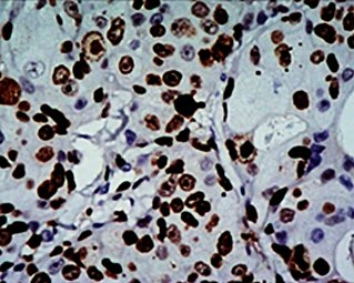 High Ki-67 expression in an invasive breast cancer, with cancer nuclei being stained (brown). There is tumor cell positivity in 70% of the cells:
Ki-67 labelling index = 70% -
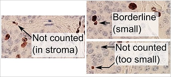 Counting positive versus negative nuclei with Ki-67 labeling, in this case in a neuroendocrine tumor of the small intestine. To count as positive, a nucleus should be at least half within the field of view, be large enough, and not be located in the stroma. Otherwise, even weakly positive nuclei count as positive.


Original Ki-67 antibody
The Ki-67 protein was originally defined by the prototype monoclonal antibody Ki-67, [17] which was generated by immunizing mice with nuclei of the Hodgkin lymphoma cell line L428. The name is derived from the city of origin ( Kiel, Germany) and the number of the original clone in the 96-well plate.
Interactions
Ki-67 (protein) has been shown to interact with CBX3. [18]
See also
- PCNA - Proliferating Cell Nuclear Antigen, expressed during the DNA synthesis.
Additional images
-
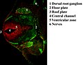 Immunofluorescent antibody staining against neurofilament (green) and Ki-67 (red) in a mouse embryo 12.5 days after fertilization. The proliferating cells are in the ventricular zone in the neural tube and therefore colored red.
 -
 Protein Ki-67 in human MCF-7 cells
 -
 Ki-67 protein (red), tubulin (green) and DNA (blue) in HeLa cells. Dividing cells show strong Ki-67 staining in cell nuclei while all cells contain large amounts of tubulin, the major component of microtubules. Antibodies, cell staining and image courtesy of EnCor Biotechnology.


References
- ^ a b c GRCh38: Ensembl release 89: ENSG00000148773 – Ensembl, May 2017
- ^ a b c GRCm38: Ensembl release 89: ENSMUSG00000031004 – Ensembl, May 2017
- ^ "Human PubMed Reference:". National Center for Biotechnology Information, U.S. National Library of Medicine.
- ^ "Mouse PubMed Reference:". National Center for Biotechnology Information, U.S. National Library of Medicine.
- ^ "Entrez Gene: Antigen identified by monoclonal antibody Ki-67".
- ^ Schonk DM, Kuijpers HJ, van Drunen E, van Dalen CH, Geurts van Kessel AH, Verheijen R, Ramaekers FC (October 1989). "Assignment of the gene(s) involved in the expression of the proliferation-related Ki-67 antigen to human chromosome 10". Hum. Genet. 83 (3): 297–9. doi: 10.1007/BF00285178. PMID 2571566. S2CID 32117144.
- ^ a b Bullwinkel J, Baron-Lühr B, Lüdemann A, Wohlenberg C, Gerdes J, Scholzen T (March 2006). "Ki-67 protein is associated with ribosomal RNA transcription in quiescent and proliferating cells". J. Cell. Physiol. 206 (3): 624–35. doi: 10.1002/jcp.20494. PMID 16206250. S2CID 22770145.
- ^ Rahmanzadeh R, Hüttmann G, Gerdes J, Scholzen T (June 2007). "Chromophore-assisted light inactivation of pKi-67 leads to inhibition of ribosomal RNA synthesis". Cell Prolif. 40 (3): 422–30. doi: 10.1111/j.1365-2184.2007.00433.x. PMC 6496591. PMID 17531085.
- ^ Sobecki M, Mrouj K, Camasses A, Parisis N, Nicolas E, Llères D, Gerbe F, Prieto S, Krasinska L, David A, others (Mar 6, 2016). "The cell proliferation antigen Ki-67 organises heterochromatin". eLife. 5 (e13722): e13722. doi: 10.7554/eLife.13722. PMC 4841783. PMID 26949251.
- ^ Scholzen T, Gerdes J (March 2000). "The Ki-67 protein: from the known and the unknown". Journal of Cellular Physiology. 182 (3): 311–22. doi: 10.1002/(SICI)1097-4652(200003)182:3<311::AID-JCP1>3.0.CO;2-9. PMID 10653597. S2CID 22944989.
- ^ Cuylen S, Blaukopf C, Politi AZ, Müller-Reichert T, Neumann B, Poser I, Ellenberg J, Hyman AA, Gerlich DW (July 2016). "Ki-67 acts as a biological surfactant to disperse mitotic chromosomes". Nature. 535 (7611): 308–12. Bibcode: 2016Natur.535..308C. doi: 10.1038/nature18610. PMC 4947524. PMID 27362226.
- ^ Bruno S, Darzynkiewicz Z (January 1992). "Cell cycle dependent expression and stability of the nuclear protein detected by Ki-67 antibody in HL-60 cells". Cell Proliferation. 25 (1): 31–40. doi: 10.1111/j.1365-2184.1992.tb01435.x. PMID 1540682. S2CID 37050410.
- ^ Darzynkiewicz Z, Zhao H, Zhang S, Lee MY, Lee EY, Zhang Z (May 2015). "Initiation and termination of DNA replication during S phase in relation to cyclins D1, E and A, p21WAF1, Cdt1 and the p12 subunit of DNA polymerase δ revealed in individual cells by cytometry". Oncotarget. 6 (14): 11735–50. doi: 10.18632/oncotarget.4149. PMC 4494901. PMID 26059433.
- ^ Sonnenblick A, Francis PA, Azim HA, de Azambuja E, Nordenskjöld B, Gutiérez J, Quinaux E, Mastropasqua MG, Ameye L, Anderson M, Lluch A, Gnant M, Goldhirsch A, Di Leo A, Barnadas A, Cortes-Funes H, Piccart M, Crown J (2015). "Final 10-year results of the Breast International Group 2-98 phase III trial and the role of Ki67 in predicting benefit of adjuvant docetaxel in patients with oestrogen receptor positive breast cancer". European Journal of Cancer. 51 (12): 1481–9. doi: 10.1016/j.ejca.2015.03.018. PMID 26074397.
- ^ Yerushalmi R, Woods R, Ravdin PM, Hayes MM, Gelmon KA (2010). "Ki67 in breast cancer: prognostic and predictive potential". The Lancet. Oncology. 11 (2): 174–83. doi: 10.1016/S1470-2045(09)70262-1. PMID 20152769.
- ^ Bánkfalvi A (November 2000). "Comparative methodological analysis of erbB-2/HER-2 gene dosage, chromosomal copy number and protein overexpression in breast carcinoma tissues for diagnostic use". Histopathology. 37 (5): 411–9. doi: 10.1046/j.1365-2559.2000.00984.x. PMID 11119122. S2CID 35267896.
- ^ Gerdes J, Schwab U, Lemke H, Stein H (1983). "Production of a mouse monoclonal antibody reactive with a human nuclear antigen associated with cell proliferation". Int. J. Cancer. 31 (1): 13–20. doi: 10.1002/ijc.2910310104. PMID 6339421. S2CID 1764319.
- ^ Kametaka A, Takagi M, Hayakawa T, Haraguchi T, Hiraoka Y, Yoneda Y (2002). "Interaction of the chromatin compaction-inducing domain (LR domain) of Ki-67 antigen with HP1 proteins". Genes to Cells. 7 (12): 1231–42. doi: 10.1046/j.1365-2443.2002.00596.x. PMID 12485163. S2CID 6802841.
External links
- Ki-67+Antigen at the U.S. National Library of Medicine Medical Subject Headings (MeSH)
- http://www.pathologyoutlines.com/topic/stainski67.html
- Overview of all the structural information available in the PDB for UniProt: P46013 (Proliferation marker protein Ki-67) at the PDBe-KB.
PDB gallery | |
|---|---|
|


| MKI67 | |||||||||||||||||||||||||||||||||||||||||||||||||||
|---|---|---|---|---|---|---|---|---|---|---|---|---|---|---|---|---|---|---|---|---|---|---|---|---|---|---|---|---|---|---|---|---|---|---|---|---|---|---|---|---|---|---|---|---|---|---|---|---|---|---|---|
|
| |||||||||||||||||||||||||||||||||||||||||||||||||||
| |||||||||||||||||||||||||||||||||||||||||||||||||||
| Identifiers | |||||||||||||||||||||||||||||||||||||||||||||||||||
| Aliases | MKI67, marker of proliferation Ki-67, KIA, MIB-, MIB-1, PPP1R105 | ||||||||||||||||||||||||||||||||||||||||||||||||||
| External IDs | OMIM: 176741 MGI: 106035 HomoloGene: 1814 GeneCards: MKI67 | ||||||||||||||||||||||||||||||||||||||||||||||||||
| |||||||||||||||||||||||||||||||||||||||||||||||||||
| |||||||||||||||||||||||||||||||||||||||||||||||||||
| |||||||||||||||||||||||||||||||||||||||||||||||||||
| |||||||||||||||||||||||||||||||||||||||||||||||||||
| Wikidata | |||||||||||||||||||||||||||||||||||||||||||||||||||
| |||||||||||||||||||||||||||||||||||||||||||||||||||
Antigen Kiel 67, also known as Ki-67 or MKI67 (marker of proliferation Kiel 67), is a protein that in humans is encoded by the MKI67 gene (antigen identified by monoclonal antibody Ki-67). [5] [6] [7]
Function
Antigen KI-67 is a nuclear protein that is associated with cellular proliferation and ribosomal RNA transcription. [7] Inactivation of antigen KI-67 leads to inhibition of ribosomal RNA synthesis, [8] but does not significantly affect cell proliferation in vivo: Ki-67 mutant mice developed normally and cells lacking Ki-67 proliferated efficiently. [9]
Use as a marker of proliferating cells
The Ki-67 protein (also known as MKI67) is a cellular marker for proliferation, [10] and can be used in immunohistochemistry. It is strictly associated with cell proliferation. During interphase, the Ki-67 antigen can be exclusively detected within the cell nucleus, whereas in mitosis most of the protein is relocated to the surface of the chromosomes. [11] Ki-67 protein is present during all active phases of the cell cycle (G1, S, G2, and mitosis), but is absent in resting (quiescent) cells (G0). [12] Cellular content of Ki-67 protein markedly increases during cell progression through S phase of the cell cycle. [13] In breast cancer Ki67 identifies a high proliferative subset of patients with ER-positive breast cancer who derive greater benefit from adjuvant chemotherapy. [14] [15]
Antibody labeling
Ki-67 is an excellent marker to determine the growth fraction of a given cell population. The fraction of Ki-67-positive tumor cells (the Ki-67 labeling index) is often correlated with the clinical course of cancer. The best-studied examples in this context are prostate, brain and breast carcinomas, as well as nephroblastoma and neuroendocrine tumors. For these types of tumors, the prognostic value for survival and tumor recurrence have repeatedly been proven in uni- and multivariate analysis.
MIB-1
Ki-67 and MIB-1 monoclonal antibodies are directed against different epitopes of the same proliferation-related antigen. Ki-67 and MIB-1 may be used on fixed sections. [16] MIB-1 is used in clinical applications to determine the Ki-67 labelling index. One of its primary advantages over the original Ki-67 antibody (and the reason why it has essentially supplanted the original antibody for clinical use) is that it can be used on formalin-fixed paraffin-embedded sections, after heat-mediated antigen retrieval (see next section below).
-
High Ki-67 expression in an invasive breast cancer, with cancer nuclei being stained (brown). There is tumor cell positivity in 70% of the cells:
Ki-67 labelling index = 70% -
Counting positive versus negative nuclei with Ki-67 labeling, in this case in a neuroendocrine tumor of the small intestine. To count as positive, a nucleus should be at least half within the field of view, be large enough, and not be located in the stroma. Otherwise, even weakly positive nuclei count as positive.
Original Ki-67 antibody
The Ki-67 protein was originally defined by the prototype monoclonal antibody Ki-67, [17] which was generated by immunizing mice with nuclei of the Hodgkin lymphoma cell line L428. The name is derived from the city of origin ( Kiel, Germany) and the number of the original clone in the 96-well plate.
Interactions
Ki-67 (protein) has been shown to interact with CBX3. [18]
See also
- PCNA - Proliferating Cell Nuclear Antigen, expressed during the DNA synthesis.
Additional images
-
Immunofluorescent antibody staining against neurofilament (green) and Ki-67 (red) in a mouse embryo 12.5 days after fertilization. The proliferating cells are in the ventricular zone in the neural tube and therefore colored red.
-
Protein Ki-67 in human MCF-7 cells
-
Ki-67 protein (red), tubulin (green) and DNA (blue) in HeLa cells. Dividing cells show strong Ki-67 staining in cell nuclei while all cells contain large amounts of tubulin, the major component of microtubules. Antibodies, cell staining and image courtesy of EnCor Biotechnology.
References
- ^ a b c GRCh38: Ensembl release 89: ENSG00000148773 – Ensembl, May 2017
- ^ a b c GRCm38: Ensembl release 89: ENSMUSG00000031004 – Ensembl, May 2017
- ^ "Human PubMed Reference:". National Center for Biotechnology Information, U.S. National Library of Medicine.
- ^ "Mouse PubMed Reference:". National Center for Biotechnology Information, U.S. National Library of Medicine.
- ^ "Entrez Gene: Antigen identified by monoclonal antibody Ki-67".
- ^ Schonk DM, Kuijpers HJ, van Drunen E, van Dalen CH, Geurts van Kessel AH, Verheijen R, Ramaekers FC (October 1989). "Assignment of the gene(s) involved in the expression of the proliferation-related Ki-67 antigen to human chromosome 10". Hum. Genet. 83 (3): 297–9. doi: 10.1007/BF00285178. PMID 2571566. S2CID 32117144.
- ^ a b Bullwinkel J, Baron-Lühr B, Lüdemann A, Wohlenberg C, Gerdes J, Scholzen T (March 2006). "Ki-67 protein is associated with ribosomal RNA transcription in quiescent and proliferating cells". J. Cell. Physiol. 206 (3): 624–35. doi: 10.1002/jcp.20494. PMID 16206250. S2CID 22770145.
- ^ Rahmanzadeh R, Hüttmann G, Gerdes J, Scholzen T (June 2007). "Chromophore-assisted light inactivation of pKi-67 leads to inhibition of ribosomal RNA synthesis". Cell Prolif. 40 (3): 422–30. doi: 10.1111/j.1365-2184.2007.00433.x. PMC 6496591. PMID 17531085.
- ^ Sobecki M, Mrouj K, Camasses A, Parisis N, Nicolas E, Llères D, Gerbe F, Prieto S, Krasinska L, David A, others (Mar 6, 2016). "The cell proliferation antigen Ki-67 organises heterochromatin". eLife. 5 (e13722): e13722. doi: 10.7554/eLife.13722. PMC 4841783. PMID 26949251.
- ^ Scholzen T, Gerdes J (March 2000). "The Ki-67 protein: from the known and the unknown". Journal of Cellular Physiology. 182 (3): 311–22. doi: 10.1002/(SICI)1097-4652(200003)182:3<311::AID-JCP1>3.0.CO;2-9. PMID 10653597. S2CID 22944989.
- ^ Cuylen S, Blaukopf C, Politi AZ, Müller-Reichert T, Neumann B, Poser I, Ellenberg J, Hyman AA, Gerlich DW (July 2016). "Ki-67 acts as a biological surfactant to disperse mitotic chromosomes". Nature. 535 (7611): 308–12. Bibcode: 2016Natur.535..308C. doi: 10.1038/nature18610. PMC 4947524. PMID 27362226.
- ^ Bruno S, Darzynkiewicz Z (January 1992). "Cell cycle dependent expression and stability of the nuclear protein detected by Ki-67 antibody in HL-60 cells". Cell Proliferation. 25 (1): 31–40. doi: 10.1111/j.1365-2184.1992.tb01435.x. PMID 1540682. S2CID 37050410.
- ^ Darzynkiewicz Z, Zhao H, Zhang S, Lee MY, Lee EY, Zhang Z (May 2015). "Initiation and termination of DNA replication during S phase in relation to cyclins D1, E and A, p21WAF1, Cdt1 and the p12 subunit of DNA polymerase δ revealed in individual cells by cytometry". Oncotarget. 6 (14): 11735–50. doi: 10.18632/oncotarget.4149. PMC 4494901. PMID 26059433.
- ^ Sonnenblick A, Francis PA, Azim HA, de Azambuja E, Nordenskjöld B, Gutiérez J, Quinaux E, Mastropasqua MG, Ameye L, Anderson M, Lluch A, Gnant M, Goldhirsch A, Di Leo A, Barnadas A, Cortes-Funes H, Piccart M, Crown J (2015). "Final 10-year results of the Breast International Group 2-98 phase III trial and the role of Ki67 in predicting benefit of adjuvant docetaxel in patients with oestrogen receptor positive breast cancer". European Journal of Cancer. 51 (12): 1481–9. doi: 10.1016/j.ejca.2015.03.018. PMID 26074397.
- ^ Yerushalmi R, Woods R, Ravdin PM, Hayes MM, Gelmon KA (2010). "Ki67 in breast cancer: prognostic and predictive potential". The Lancet. Oncology. 11 (2): 174–83. doi: 10.1016/S1470-2045(09)70262-1. PMID 20152769.
- ^ Bánkfalvi A (November 2000). "Comparative methodological analysis of erbB-2/HER-2 gene dosage, chromosomal copy number and protein overexpression in breast carcinoma tissues for diagnostic use". Histopathology. 37 (5): 411–9. doi: 10.1046/j.1365-2559.2000.00984.x. PMID 11119122. S2CID 35267896.
- ^ Gerdes J, Schwab U, Lemke H, Stein H (1983). "Production of a mouse monoclonal antibody reactive with a human nuclear antigen associated with cell proliferation". Int. J. Cancer. 31 (1): 13–20. doi: 10.1002/ijc.2910310104. PMID 6339421. S2CID 1764319.
- ^ Kametaka A, Takagi M, Hayakawa T, Haraguchi T, Hiraoka Y, Yoneda Y (2002). "Interaction of the chromatin compaction-inducing domain (LR domain) of Ki-67 antigen with HP1 proteins". Genes to Cells. 7 (12): 1231–42. doi: 10.1046/j.1365-2443.2002.00596.x. PMID 12485163. S2CID 6802841.
External links
- Ki-67+Antigen at the U.S. National Library of Medicine Medical Subject Headings (MeSH)
- http://www.pathologyoutlines.com/topic/stainski67.html
- Overview of all the structural information available in the PDB for UniProt: P46013 (Proliferation marker protein Ki-67) at the PDBe-KB.
PDB gallery | |
|---|---|
|