Original file (1,024 × 768 pixels, file size: 415 KB, MIME type: image/png)
| This is a file from the
Wikimedia Commons. Information from its
description page there is shown below. Commons is a freely licensed media file repository. You can help. |
Summary
| Description |
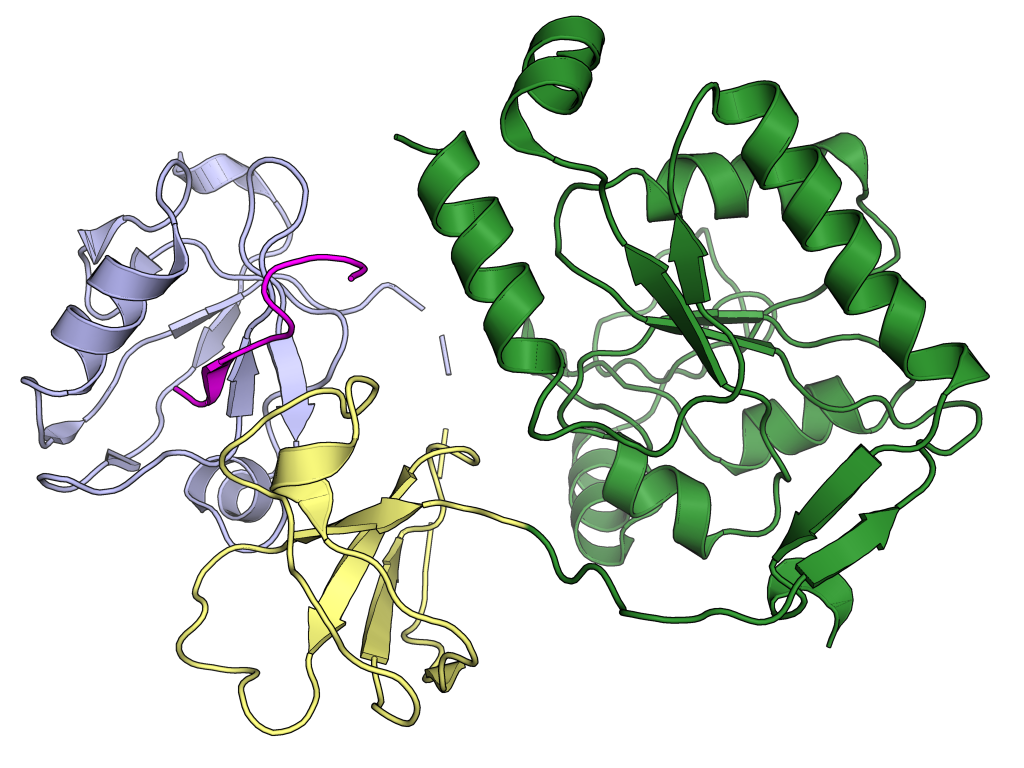
Cryo-electron microscopy structure showing a peptide derived from the the SARS-CoV-2 envelope protein PDZ-binding domain (magenta) in complex with a construct containing the PDZ (blue), SH3 (yellow), and guanylate kinase-like (GK, green) domains from a host cell protein, human PALS1 (uniprot Q8N3R9). Rendered with PyMol from PDB: 7M4R chains A and C. Structural basis for SARS-CoV-2 envelope protein recognition of human cell junction protein PALS1 Jin Chai, Yuanheng Cai, Changxu Pang, Liguo Wang, Sean McSweeney, John Shanklin, Qun Liu Nature Communications volume 12, Article number: 3433 (2021) DOI: 10.1038/s41467-021-23533-x |
| Date | |
| Source | Own work |
| Author | Opabinia regalis |
Licensing
- You are free:
- to share – to copy, distribute and transmit the work
- to remix – to adapt the work
- Under the following conditions:
- attribution – You must give appropriate credit, provide a link to the license, and indicate if changes were made. You may do so in any reasonable manner, but not in any way that suggests the licensor endorses you or your use.
- share alike – If you remix, transform, or build upon the material, you must distribute your contributions under the same or compatible license as the original.

|
Permission is granted to copy, distribute and/or modify this document under the terms of the GNU Free Documentation License, Version 1.2 or any later version published by the Free Software Foundation; with no Invariant Sections, no Front-Cover Texts, and no Back-Cover Texts. A copy of the license is included in the section entitled GNU Free Documentation License. |
File history
Click on a date/time to view the file as it appeared at that time.
| Date/Time | Thumbnail | Dimensions | User | Comment | |
|---|---|---|---|---|---|
| current | 02:05, 29 June 2021 |
| 1,024 × 768 (415 KB) | Opabinia regalis | {{Information |Description=Cryo-electron microscopy structure showing a peptide derived from the the SARS-CoV-2 envelope protein PDZ-binding domain (magenta) in complex with a construct containing the PDZ (blue), SH3 (yellow), and guanylate kinase-like (GK, green) domains from a host cell protein, human PALS1 (uniprot Q8N3R9). Rendered with PyMol from {{PDB|7M4R}} chains A and C. Structural basis for SARS-CoV-2 envelope protein recognition of human cell junction protein PALS1 Jin Chai, Yuan... |
File usage
Global file usage
The following other wikis use this file:
- Usage on ar.wikipedia.org
Metadata
Original file (1,024 × 768 pixels, file size: 415 KB, MIME type: image/png)
| This is a file from the
Wikimedia Commons. Information from its
description page there is shown below. Commons is a freely licensed media file repository. You can help. |
Summary
| Description |
Cryo-electron microscopy structure showing a peptide derived from the the SARS-CoV-2 envelope protein PDZ-binding domain (magenta) in complex with a construct containing the PDZ (blue), SH3 (yellow), and guanylate kinase-like (GK, green) domains from a host cell protein, human PALS1 (uniprot Q8N3R9). Rendered with PyMol from PDB: 7M4R chains A and C. Structural basis for SARS-CoV-2 envelope protein recognition of human cell junction protein PALS1 Jin Chai, Yuanheng Cai, Changxu Pang, Liguo Wang, Sean McSweeney, John Shanklin, Qun Liu Nature Communications volume 12, Article number: 3433 (2021) DOI: 10.1038/s41467-021-23533-x |
| Date | |
| Source | Own work |
| Author | Opabinia regalis |
Licensing
- You are free:
- to share – to copy, distribute and transmit the work
- to remix – to adapt the work
- Under the following conditions:
- attribution – You must give appropriate credit, provide a link to the license, and indicate if changes were made. You may do so in any reasonable manner, but not in any way that suggests the licensor endorses you or your use.
- share alike – If you remix, transform, or build upon the material, you must distribute your contributions under the same or compatible license as the original.
|
|
Permission is granted to copy, distribute and/or modify this document under the terms of the GNU Free Documentation License, Version 1.2 or any later version published by the Free Software Foundation; with no Invariant Sections, no Front-Cover Texts, and no Back-Cover Texts. A copy of the license is included in the section entitled GNU Free Documentation License. |
File history
Click on a date/time to view the file as it appeared at that time.
| Date/Time | Thumbnail | Dimensions | User | Comment | |
|---|---|---|---|---|---|
| current | 02:05, 29 June 2021 |
| 1,024 × 768 (415 KB) | Opabinia regalis | {{Information |Description=Cryo-electron microscopy structure showing a peptide derived from the the SARS-CoV-2 envelope protein PDZ-binding domain (magenta) in complex with a construct containing the PDZ (blue), SH3 (yellow), and guanylate kinase-like (GK, green) domains from a host cell protein, human PALS1 (uniprot Q8N3R9). Rendered with PyMol from {{PDB|7M4R}} chains A and C. Structural basis for SARS-CoV-2 envelope protein recognition of human cell junction protein PALS1 Jin Chai, Yuan... |
File usage
Global file usage
The following other wikis use this file:
- Usage on ar.wikipedia.org