Vector-borne diseases are human infections caused by
bacteria,
parasites, and
viruses that are spread by
vectors.[1] A vector is a living
organism that spreads
pathogens; in other words, it carries disease-causing organisms from one host to another. Vectors are normally considered to be
invertebrate animals, usually
arthropods, but also include
rodents, which carry the infectious disease from a reservoir to a susceptible host.[2] Vectors of human disease are typically a species of
mosquitoes,
ticks,
flies, and
fleas that are able to transmit bacteria, viruses, or parasites to humans and other vertebrate hosts.[3] A disease that is transmitted to humans, plants, or animals by any agent, arthropod, or fomite is a vector-borne disease.[4]
When trying to determine what the deadliest animal is in the world, a mosquito is likely not the first thing that comes to mind. However, the ability of mosquitoes to transmit disease causes millions of deaths each year. Female mosquitoes feed on the blood of infected animals and then spread the disease to humans through blood feeding. The diseases that they carry include malaria, dengue, yellow fever and countless others. There are treatments for some of these diseases, but for many there still is not treatment beyond supportive care. With the changing environmental climate, the concern about mosquito-borne disease becomes more serious with increases in flood zones and stagnant water. Preventative strategies include vaccinations when possible, wearing clothing to cover exposed areas, the use of insect repellent and destroying areas of breeding grounds.
Aedes Mosquito
Aedes Mosquito
Chikungunya
Vector type: Aedes aegypti and Aedes albopictus. This disease is found transmitted by these mosquito types in the Americas, Europe, Africa, Asia and India. When the breeding sites are close to homes they become a significant risk factor.[5]
Transmission: Chikungunya is transmitted via the bite of an infected female mosquito. These mosquitoes bite most frequently during the daylight, with peak activity occurring during the dawn and dusk. Recorded outbreaks are usually separated by period of 10 years.[6]
Signs and Symptoms: The onset of signs and symptoms of Chikungunya occur between 4-8 days and last for a duration of 2-3 days. The primary symptoms are fever with severe joint and muscle pain, headache, nausea, fatigue and rash. The name Chikyngunya means "to be contorted" because of the joint pain.[7] Most patients are able to fully recover but there are some serious complications that can occur. These complications involve the eyes, brain and heart and can cause death in older individuals. The signs and symptoms can often be confused with Dengue fever.
Diagnosis: Chikungunya is diagnosed with the use of serological tests. After infection, there is lifelong immunity against the disease.
Treatment: The most common ways to treat Chikungunya is with the use of supportive medicine. According to the WHO, this includes fluids, pain medicines and analgesics.[8] Unfortunately, there are currently no commercial vaccines available.
Prevention: The greatest preventative measures against the spread of Chikungunya involve mosquito protection. This can be done through the use of repellents, protective clothing, mosquito netting, window screens and breeding ground destruction.
Dengue Fever
Vector type: Aedes aegypti; There are 4 distinct serotypes of the Dengue virus that fall under that viral type of Flavivirus, Flaviviridae.[9]
Transmission: The disease is common in urban poor areas, suburbs and rural settings. The incidence has increased 30 times in the last 50 years, putting almost half of the worlds population at risk.[10]
Signs and Symptoms: Dengue presents with severe flu-like signs and symptoms and is at times referred to as "break-bone" fever. The onset of symptoms takes 4-10 days and lasts from 2-7 days. There is a severe headache, pain behind the eyes, nausea/vomiting, swollen glands, muscle and joint pains, rash, and skin lesions. Severe Dengue can also occur due to plasma leaking which causes an accumulation of fluid leading to respiratory distress and severe bleeding. This severe form of the disease is also known as dengue hemorrhagic fever. According to the WHO it and is one of the leading causes of death in Asian and Latin American countries. To detect the severe form of the disease patients should be monitored to see if after 3-7 days there is a drop in temperature. This is accompanied by severe abdominal pain, persistent vomiting, rapid breathing, bleeding gums, bloody vomit, fatigue and restlessness. A patient that recovers from the illness develops lifelong immunity from the specific serotype, but only partial from the other three infectious serotypes.[11]
Diagnosis: Dengue is detected via dengue NS1 serological testing.
Treatment: There is no specific treatment for Dengue. It is best to seek medical advice, rest and fluids. To treat the symptoms, paracetamol can be used to treat the fever and joint pains. However, it is important to avoid the use of aspirin or ibuprofen to decrease the bleeding risk.
Prevention: To prevent Dengue, it is important to follow mosquito protocol. It is also important to employ solid waste disposal and water storage practices.
Lymphatic Filariasis
also known as "elephantiasis"
Vector types: Culex in widespread urban and semi-urban areas. Anopheles in rural areas and Aedes in endemic islands in the Pacific.
Transmission: Lymphatic filiariasis is caused by an infection with nematodes (roundworms). The main types are Wuchereria bancrofti (90%) of cases, Brugia malaui and Brugia timori. Mosquitoes are infected with the microfilariae, which are then passed on to humans. The adults works then lodge into the lymphatic vessels and cause severe damage and lymphodema. This is usually acquired in childhood and leads to permanent disability.
Signs and Symptoms: There are three main divisions of the signs and symptoms associated with lymphatic filiariasis; asymptomatic, chronic and acute. In asymptomic infections, there are no external signs and symptoms of infection. However, there can still be some internal damage, especially to the lymphatics and kidneys. In chronic lymphatic filiariasis, there is lymphedema (tissue swelling), elephantiasis (skin/tissue thickening) of limbs and scrotal swelling (hydrocele). There can also be some involvement of the breasts and genitals.[12] The largest concern in those with chronic lymphatic filiariasis lies in the social stigma that follows. There is often severe stigma and physical disability that occurs as a results of this infection that perpetuates socioeconomic burdens, feelings of isolation and poverty. The last type of infection is acute lymphatic filiariasis. The acute infection has local inflammation of the skin, lymph nodes and lymphatic vessels. This can then lead to chronic lymphoedema or elephantiasis that can last for weeks. This occurs because of the immune response to the parasite and/or secondary bacterial skin infection due to the poor lymphatic drainage.
Treatment: The best treatment is through the use of preventative drug programs. This is done with a combined dose of albendazole+ivermetin or DEC to reduce the amount of microfiliariae.[13] By reducing the density of the parasite when it is small, the blockage of lymphatics by the adult worms can be prevented. It is important to note that this drug combination is ineffective against the adult forms. Other treatments include the relief of symptoms such as surgery for hydroceles.
Prevention: The WHO has implemented the Global Programme to Eliminate Lymphatic Filiariases (GPELF) in 2000.[14] The purpose of this program is to facilitate large scale annual treatments of preventative drug regimines in high risk areas. The program also includes increased management of those with disabilities resulting from infection with the end goal of decrease the stigma and socioeconomic burdens on these individuals. Mosquito protocol with insecticide treated nets, indoor spraying and personal protection measures are also encouraged.
Rift Valley Fever
Vector Type: Aedes and Culex mosquitoes.
Transmission: Rift Valley Fever occurs most commonly with direct/indirect contact with the blood or organs of infected animals. However, transmission can also occur if a mosquito is infected by feeding on an infected animal. Those who are most at risk include occupations like vets, herders, farmers and butchers. Transmission can also occur through inoculation in an open would or through inoculation.[15]
Signs and Symptoms: The onset of Rift Valley Fever symptoms occur between 2-6 days. Under normal conditions, it presents with flu-like signs and symptoms, fever, muscle and joint pain and headache. There have also been reports of neck stiffness, sensitivity to light, loss of appetite and vomiting. The signs and symptoms may be confused with those of meningitis. There is also a severe form of the disease that can take on three different forms. The first includes retinal lesions of the eyes. The second causes meninogoencephalitis with a presentation of intense headache, loss of memory, hallucinations, confusion, convulsions and even coma. These do not also appear immediately with onset of symptoms, but can occur more than 60 days post-infection. Lastly, there is a form associated with hemorrhagic fever. This occurs 2-4 days following the initial onset of symptoms. There is severe damage to the liver with jaundice/yellowing, vomiting blood, bleeding from the gums and nose and death between 3-6 days.[16]
Diagnosis: Rift Valley Fever can be diagnosed with RT-PCR assay, ELISA or viral isolation of cell culture
Treatment: There is no specified treatment, and medical intervention is supportive care.
Prevention: There is no commercially available vaccine, but there is an inactivated vaccine that can be used for at risk animals. It is best to closely monitor livestock in high risk areas to be on the lookout for outbreak.[17] It is also important to reduce exposure in at risk occupations and ensuring proper cooking of blood, meat and milk products. Lastly, mosquito protection is also important, especially in high risk seasons like following the rainy season.
Yellow Fever
Vector Type: Aedes mosquitoes (domestic, wild and semi-domestic forms)
Geography: endemic to Africa, Central and South America
Transmission: There are three types of transmission cycles associated with Yellow Fever; Jungle, Intermediate and Urban.[18] In the Jungle transmission cycle, the virus is transferred from monkeys to mosquitoes to humans. In the Intermediate cycle, the virus is spread by semi-domestic breeds of Aedes mosquitoes that are infected by monkeys and then infect humans through blood feeding. Due to increased contact between villages and areas of infection, there is increased transmission by this cycle, making is the most common type of outbreak in Africa. This also means that there can be outbreaks of disease in multiple villages occurring at the same time. Lastly, is the urban transmission cycle which is responsible for large epidemics in densely populated regions.[19]
Signs and Symptoms: Yellow Fever is an acute viral hemorrhagic disease. The associated symptoms unique to this disease include fever, headache, jaundice, muscle pain, nausea and vomiting, fatigue and black vomit. Most signs and symptoms only last 3-4 days. However, some may become severe cases which have a 50% mortality rate. The severe form occurs within the first 24 hours after it looks like recovery is taking place. The severe signs and symptoms present with a return of a high fever, severe jaundice with yellowing of the eyes and skin due to kidney and liver damage, dark urine and bleeding. This is a biphasic disease with the initial symptoms resulting from damage to the vascular endothelium, this then progresses to the liver during later replication.[18]
Diagnosis: Yellow Fever can be misdiagnosed as either severe malaria, viral hepatitis, poisoning or other types of hemorrhagic fevers when it is in the early phases. PCR and ELISA assays can be run to diagnose. A liver biopsy can also be done, you would expect to see Councilman bodies in the cytoplasm (esophinophilic hyaline masses).[19]
Treatment: There is no specific anti-viral drug therapy for infection with yellow fever. The recommended supportive treatment includes care for dehydration, fever and the complications from liver and kidney failure. Antibiotics may also be used to prevent any associated bacterial infections.
Prevention: There is a very successful vaccine that confers lifelong protection with a single dose and no need for a booster. The vaccine is an attenuated vaccine strain 17D, which is 99% effective within 30 days. [18]The vaccine is recommended in travelers to any endemic regions, in infants and even in the use of mass vaccination campaigns. It is also important to maintain mosquito protection protocol, added larvicides to water containers or any sites of stagnant water and routine insecticide spraying. Lastly, is maintaining epidemic preparedness. This results in fast detection with treatment and prevention response, which is crucial for controlling any outbreaks.
Zika
Vector Type: Aedes aegypti
Transmission: Zika is transmitted from the bite of an infected female mosquito when it feeds on humans. The most frequent times for infection occur during the day, especially at dawn and dusk when mosquitoes are the most prevalent. Sexual contact has also been found to be a pathway for transmission of Zika. [20]
Signs and Symptoms: Those infected present with a mild fever, skin rash, muscle and joint pain, headache or conjunctivitis that typically lasts for 2-7 days. The key features of Zika infection include the neurological components of microcephaly in pregnant women that is manifest in the fetus and Guilian-Barre syndrome with resulting transient paralysis.[21] Otherwise, it presents in a manner similar to that of the other arboviruses like Dengue.
Diagnosis: Zika can be detected through lab testing of blood, urine, saliva or semen.
Treatment: The recommended treatment options are largely supportive with rest, fluids and controlling pain and fever with normal drug regimens.
Prevention: There is no vaccine currently available for Zika. In order to protect against mosquito bites is most important with physical barriers like nets or covered clothing. Insect repellent that contains DEET, IR3535 or icaridin are other methods to prevent mosquitoes from feeding.[22] It is also important to detect and remove mosquito breeding sites. In terms of sexual transmission, safe sex practices and barriers like condoms are recommended, especially in women who are pregnant or may become pregnant.
Anopheles Mosquito
Anopheles Mosquito
Malaria
Vector Type: Anopheles species
Transmission: Malaria is transmitted via infection of a female mosquito with the plasmodium parasites; P. falciparum, P. vivax, P. ovale and P. malariae. According to the WHO, Africa carries the highest burden of disease with 90% of malaria cases and 91% of malarial deaths. Malaria infection is especially prevalent after the rainy season.[23] Those that are most at risk for becoming infected include infants, children, pregnant women and those with HIV/AIDS.
Signs and Symptoms: Malaria is an acute febrile illness that has an onset of symptoms that occur around 10-15 days after infection. The signs and symptoms include fever, headache and splenomegaly. The severity of disease is dependent on the type of plasmodium parasite.
P. Falciparum: The most severe form of the disease occurs with the plasmodium falciparum species that has a distinctive irregular fever pattern. In children, severe malaria can lead to severe anemia, respiratory distress as a result of metabolic acidosis and cerebral malaria. In adults, there can be multi-organ involvement and failure. The cause of these symptoms lies in the occlusion of capillaries by the parasitized red blood cells.[24]
P. vivax and P. ovale: These forms of disease are characterized by a fever pattern that has a 48 hour cycle with a fever occurring on the first and third days. There is a dormant form of these infections resulting from the hypnozoites in the liver.
P. malariae: This form of infection has a quartan fever pattern occuring every four days.
Diagnosis: Microscopy or rapid diagnostic testing can be done. P. falciparum can be diagnosed with the use of Giemsa staining with multiple ring forms and crescents seen. P. vivax and P. ovale can be diagnosed via Giemsa staining with red granules called Schuffner stippling throughout the red blood cell cytoplasm. P. malariae can be diagnosed through Giemsa staining with bar and band forms present.[19]
Treatment: The treatment of malaria can be complicated and depends on the type of infection and severity of disease. P. falciparum is treated with chloroquine and resistant strains are treated with quinine sulfate. P. vivas and ovale are treated with chloroquine and primaquine for the dormant liver form. Mefloquine or atovaquonone/proguanil can also be used to treat resistant strains. If the illness becomes life-threatening then IV quinidine or artesunate should be used, while making sure to test of G6PD deficiency.[19]
Prevention: Anti-malarial drugs and chemoprophylaxis are the most important in preventing the disease. Insecticide mosquito nets, indoor spraying with insecticides and disease surveillance are also important preventative measures. There is a vaccine available called Mosquirix that might provide some partial protection in young children.[24]
Lymphatic Filariasis
also known as "elephantiasis"
Vector types: Culex in widespread urban and semi-urban areas. Anopheles in rural areas and Aedes in endemic islands in the Pacific.
Transmission: Lymphatic filiariasis is caused by an infection with nematodes (roundworms). The main types are Wuchereria bancrofti (90%) of cases, Brugia malaui and Brugia timori. Mosquitoes are infected with the microfilariae, which are then passed on to humans. The adults works then lodge into the lymphatic vessels and cause severe damage and lymphodema. This is usually acquired in childhood and leads to permanent disability.
Signs and Symptoms: There are three main divisions of the signs and symptoms associated with lymphatic filiariasis; asymptomatic, chronic and acute. In asymptomic infections, there are no external signs and symptoms of infection. However, there can still be some internal damage, especially to the lymphatics and kidneys. In chronic lymphatic filiariasis, there is lymphedema (tissue swelling), elephantiasis (skin/tissue thickening) of limbs and scrotal swelling (hydrocele). There can also be some involvement of the breasts and genitals.[12] The largest concern in those with chronic lymphatic filiariasis lies in the social stigma that follows. There is often severe stigma and physical disability that occurs as a results of this infection that perpetuates socioeconomic burdens, feelings of isolation and poverty. The last type of infection is acute lymphatic filiariasis. The acute infection has local inflammation of the skin, lymph nodes and lymphatic vessels. This can then lead to chronic lymphoedema or elephantiasis that can last for weeks. This occurs because of the immune response to the parasite and/or secondary bacterial skin infection due to the poor lymphatic drainage.
Treatment: The best treatment is through the use of preventative drug programs. This is done with a combined dose of albendazole+ivermetin or DEC to reduce the amount of microfiliariae.[13] By reducing the density of the parasite when it is small, the blockage of lymphatics by the adult worms can be prevented. It is important to note that this drug combination is ineffective against the adult forms. Other treatments include the relief of symptoms such as surgery for hydroceles.
Prevention: The WHO has implemented the Global Programme to Eliminate Lymphatic Filiariases (GPELF) in 2000.[14] The purpose of this program is to facilitate large scale annual treatments of preventative drug regimines in high risk areas. The program also includes increased management of those with disabilities resulting from infection with the end goal of decrease the stigma and socioeconomic burdens on these individuals. Mosquito protocol with insecticide treated nets, indoor spraying and personal protection measures are also encouraged.
Culex Mosquito
Culex Mosquito
Japanese Encephalitis
Vector Type: Culex Species
Transmission: The transmission cycle includes mosquitoes, pigs, water birds and humans. Infection is particularly high in the warm, rainy and periods before the rice harvest. According to the WHO, Japanese encephalitis is the main cause of viral encephalitis in Asia with outbreaks occurring at intervals of 2-15 years.[25] Rural and semi-urban regions are the ones that are most prone to outbreak.
Signs and Symptoms: The most often types of signs and symptoms for Japanese encephalitis include a mild headache and fever. However, a severe form of the disease can occur. The presentation of the severe form of the disease includes a fast onset of high fever, headache, neck stiffness, disorientation, seizures, spastic paralysis and death. According to the WHO, the fatality rate can be as high as 30%. Of those that survive the severe form of JEV, 20-30% suffer permanent intellectual and neurological complications like paralysis, recurrent seizures and inability to speak.[26]
Diagnosis: lab testing of serum or cerebrospinal fluid is used if possible to obtain.
Treatment: There is no cure for Japanese Encephalitis. Intervention consists of relieving the clinical signs through supportive care.
Prevention: In addition to mosquito protocols, there are vaccines that can be used as parts of national immunization schedules. There are currently four types of vaccines available; inactivated mouse brain derived vaccines, inactivated Vero cell derived vaccines, live attenuated vaccines and live recombinant vaccines.[27]
Lymphatic Filariasis
also known as "elephantiasis"
Vector types: Culex in widespread urban and semi-urban areas. Anopheles in rural areas and Aedes in endemic islands in the Pacific.
Transmission: Lymphatic filiariasis is caused by an infection with nematodes (roundworms). The main types are Wuchereria bancrofti (90%) of cases, Brugia malaui and Brugia timori. Mosquitoes are infected with the microfilariae, which are then passed on to humans. The adults works then lodge into the lymphatic vessels and cause severe damage and lymphodema. This is usually acquired in childhood and leads to permanent disability.
Signs and Symptoms: There are three main divisions of the signs and symptoms associated with lymphatic filiariasis; asymptomatic, chronic and acute. In asymptomic infections, there are no external signs and symptoms of infection. However, there can still be some internal damage, especially to the lymphatics and kidneys. In chronic lymphatic filiariasis, there is lymphedema (tissue swelling), elephantiasis (skin/tissue thickening) of limbs and scrotal swelling (hydrocele). There can also be some involvement of the breasts and genitals.[12] The largest concern in those with chronic lymphatic filiariasis lies in the social stigma that follows. There is often severe stigma and physical disability that occurs as a results of this infection that perpetuates socioeconomic burdens, feelings of isolation and poverty. The last type of infection is acute lymphatic filiariasis. The acute infection has local inflammation of the skin, lymph nodes and lymphatic vessels. This can then lead to chronic lymphoedema or elephantiasis that can last for weeks. This occurs because of the immune response to the parasite and/or secondary bacterial skin infection due to the poor lymphatic drainage.
Treatment: The best treatment is through the use of preventative drug programs. This is done with a combined dose of albendazole+ivermetin or DEC to reduce the amount of microfiliariae.[13] By reducing the density of the parasite when it is small, the blockage of lymphatics by the adult worms can be prevented. It is important to note that this drug combination is ineffective against the adult forms. Other treatments include the relief of symptoms such as surgery for hydroceles.
Prevention: The WHO has implemented the Global Programme to Eliminate Lymphatic Filiariases (GPELF) in 2000.[14] The purpose of this program is to facilitate large scale annual treatments of preventative drug regimines in high risk areas. The program also includes increased management of those with disabilities resulting from infection with the end goal of decrease the stigma and socioeconomic burdens on these individuals. Mosquito protocol with insecticide treated nets, indoor spraying and personal protection measures are also encouraged.
West Nile Fever
Vector Type: Culex (species Pipiens)
Transmission: West Nile Fever is present in Africa, Europe, Middle East, North America and West Asia. The transmission cycle is between birds (crows and jay as natural hosts) and mosquitoes primarily, but also humans and horses.[28] There is also a very small percentage of infection from organ transplants, blood transfusions and breast milk.
Signs and Symptoms: 80% of West Nile infections are asymptomatic. However, for the other 20% there can be fevers, head and body aches, nausea, skin rash and swollen lymph glands. The incubation period before the onset of symptoms ranges from 3-14 days. There is also a severe form of the disease that has a neuroinvasive component. This presents with meningitis, high fever, neck stiffness, disorientation, convulsions, muscle weakness and paralysis. Those who are over 50 years old are most at risk. West Nile is the leading cause of arboviral encephalitis in the United States.[29]
Diagnosis: IgG antibody sero-conversion, ELISA-IgM, neutralization assays, PCR and viral isolation by cell culture is used to detect West Nile Fever.[19] IgM can also be detected in CSF and serum.
Treatment: Supportive care is recommended. For those with the neuroinvasive form of the disease, hospitalization with the use of IV fluids, respiratory support and antibiotics to prevent additional infection is recommended.[30]
Prevention: There are no vaccines for West Nile for humans, but ones that can be used for horses and to prevent transmission in horses.[31] An active animal health surveillance system can also be used to monitor outbreaks. Mosquito prevention is the most widely and effective use of prevention of infection.
Adult Deer Tick
Vector Type: Ticks Information
Ticks are the most significant vector of disease amongst wild and domestic animals in the world and are only second to mosquitoes in transmitting disease to humans.[32] In the United States, ticks are the leading cause of vector-borne infectious disease.[33]Ticks are external parasites that have been associated with the transmission of
pathogens for centuries dating back to the ancient times.[34] For ticks to complete their life-cycle, they are dependent on wild animals like deer and rodents for nourishment and transportation but also feed on domestic animals as hosts. There are two main families of ticks:
Ixodidae (
hard ticks) and
Argadidae (
soft ticks), that transmit disease to humans.[35] Hard ticks have a tough shield protecting their back and are darker in color while soft ticks do not have a protective shield, are lighter in color, and appear leathery.[36] Hard ticks present a substantial health risk and spread a majority of all
tick-borne infectious disease important to humans caused by bacteria, protozoa, and viruses. The life cycle of hard ticks is usually around two years during which they go through four growth phases: egg, larva, nymph, and adult.[37] In order for ticks to feed they must climb onto hosts (they do not jump or fly). One method to accomplish this is for ticks to wait for a host to pass by while sitting in a “questing” position characterized by grasping vegetation with their back legs and holding their front two legs out. When a host brushes by they quickly climb on and find a suitable place to feed.[38] Ticks can live in grasslands, wooded areas, wet or dry climates, and relatively temperate regions around the world in both North and South America, Africa, Europe, Asia, and Australia.
Crimean-Congo Hemorrhagic Fever (CCHF) is a widespread tick-borne disease caused by the transmission of Nairovirus which leads to viral hemorrhagic fever eruptions in humans. CCHF has a case-fatality rate of 10-40% and a mortality rate of 30%.[39] It is prevalent in the Middle East, Asia, the Balkins, Africa, and other countries south of the 50th parallel north.[40] Hosts of bunyaviridae ticks include domestic and wild animals such as cattle, sheep, goats, and ostriches in endemic areas.
Transmission: Crimean-Congo Hemorrhagic Fever (CCHF) is primarily transmitted to humans by tick bites or infected livestock animals.[41] Close contact with infected animal tissue immediately following the harvesting/slaughter of infected animals can cause infection in humans. Human-to-human transmission is possible with close contact to infected blood, secretions, organs, and bodily fluids. Hospital-acquired infections may happen due to unsanitary hygiene practices as well. Most CCHF infections occur in the livestock industry such as agricultural workers, veterinarians, and slaughterhouse workers.[42]
CCHF Skin LesionsSigns and symptoms: The incubation period for CCHF depends on how the Nairovirus was transmitted. Tick bite infections have an incubation period of 1-3 days (maximum of 9 days) before symptoms will show.[43] Infections caused by exposure to infected tissues or blood have an incubation period of 5-6 days (maximum of 13 days).[44] Symptoms of CCHF are sudden and may include fever, muscle aches, neck pain/stiffness, dizziness, headaches, sore eyes, backache, sensitivity to light, nausea, vomiting, sore throat, diarrhea, abdominal pain, and mood swings.[45] After 2-4 days symptoms may be replaced by sleepiness and depression and the abdominal pain may focus on the upper right abdominal area with detectable liver enlargement.[46] Clinical signs include fast heart rate, enlarged lymph nodes, and rashes in the mouth and throat as well as on the skin. Severely ill patients may progress into hepatitis and kidney, liver, and pulmonary failure after the fifth day. In severe cases, death usually occurs in the second week of infection.[47]
Diagnosis: CCHF can be diagnosed by a variety of laboratory tests including: enzyme-linked immunosorbent assay (ELISA), antigen detection, serum neutralization, reverse transcriptase polymerase chain reaction (RT-PCR) assay; and virus isolation by cell culture.[48]
Treatment: There is no absolute cure for CCHF. Treatment is achieved through supportive care for associated symptoms. An antiviral drug called ribavirin has be used to treat CCHF with notable benefit but lacks scientific evidence.[49]
Prevention: Controlling tick-borne infections like CCHF are difficult because of the level of difficulty in breaking the tick-animal-tick chain of infection (sequential infections; one after another). There are no current animal vaccinations and no clinically approved human vaccinations to prevent CCHF.[50] Raising awareness, using acaricides (chemicals to kill ticks), and personal protective equipment (long sleeves and pants, chemically treated clothing, skin repellent, light colored clothing to enhance visibility, gloves when handling livestock and tissues) are the best prevention methods to reduce infection.[51]
Lyme disease is the leading vector-borne disease in the United States with an estimated 329,000 cases diagnosed annually and one of the fastest growing vector-borne diseases in both the United States and Western Europe.[52] It was first identified in the United States in the mid-1970's during an investigation into an arthritis epidemic in Old Lyme, Connecticut.[53] Lyme Disease is caused by the bite of a deer tick which transmits the bacteria
borrelia burgdorferi. If untreated, Lyme disease can lead to debilitating illness affecting the joints, heart, brain, and nervous system.[54] Lyme disease predominately located in the Northeastern and upper Midwestern United States and Western Europe but can be found in the Pacific coast, Mid-Atlantic coast, and other parts of Europe.[55]
Transmission: Lyme Disease is spread by infected ixodes ticks when they bite hosts. In order for Lyme disease to be transmitted the tick must feed for a period of between 36-38 hours.[56] Ticks usually choose feeding areas in hard to detect places such as the groin, armpits, and scalp.[57] Most humans are infected by ticks in the nymph life stage due to their small size and difficulty to spot but, adult ticks may also infect humans as well.[58] Ixodes ticks feed on animals (most notably whitetail deer) and domestic pets like dogs and cats who then bring them into human domiciles where they can then climb onto humans. Wild and domestic animals can contract Lyme disease from infected ticks but there is no evidence that infected animals can transmit the disease to humans.[59] The incubation period for Lyme disease can range from anywhere between a few days up to a month (average time is 7 days) before the characteristic skin lesions appear.[60]
Erythema Migrans Skin LesionSigns and symptoms: The most notable sign of Lyme disease are skin lesions known as
“erythema migrans” (Latin for migrating redness) which occur in 90% of cases.[61][62] The skin lesions begin at the site of the bite and gradually expand up to a 12inch area.[63] Other associated symptoms are arthritis, headache, fatigue, fever, chills, muscle and joint soreness, and in later stages facial paralysis, heart palpitations, dizziness, shortness of breath, numbness and tingling in extremities, and memory loss.[64][65] End stages of the disease can lead to cranial neuritis, meningitis, and Bell’s palsy.
Diagnosis: Lyme disease can be diagnosed based on signs and symptoms with a history of exposure to endemic ixodes tick areas and laboratory tests for positive cultures of the borrelia burgdorferi bacteria.[66][67]
Treatment: Early stage treatment includes the administration of doxycycline, amoxicillin, cefuroxime, and azithromycin taken orally over a 2-3 week period.[68] Late stage treatment usually requires intravenous administration of penicillin or ceftriaxone over a 2-3 week period.[69]
Prevention: Reducing exposure to ticks is the best prevention method.[70] After exposure to tick endemic areas, thorough self-check procedures are recommended to reduce possibility of infection. Other preventive methods include wearing long sleeves and pants, chemically treat clothing with anti-tick chemicals, application of tick repellent to skin and probable feeding areas, and avoidance of contact with wildlife in endemic regions.
Tick-borne
Relapsing Fever (TBRF) is a bacterial infection that results in reoccurring (relapsing) fever, headache, muscle and joint pain, and nausea.[71] It is caused by
Borrelia bacteria which is transmitted through the bite of an
ornithodoros tick.[72] TBRF was first discovered in the United States in 1915.[73] The primary host of
ornithodoros ticks are rodents vice deer and domestic animals. Common hosts of these soft ticks are ground squirrels, chipmunks, prairie dogs, mice, and owls.[74] Soft ticks do not search for prey by “questing” in grasslands and wooded areas, rather they live in rodent burrows where they feed at will while rodents sleep and can survive and remain infectious for several years without feeding.[75][76] The average lifespan of soft ticks is around 10 years but can survive up to 20 years.[77] Most infections occur in the spring and summer months when humans vacation in mountainous rustic rodent infested cabins. TBRF is distributed throughout mountainous and plateau regions of North America, Mexico, South and Central America, the Mediterranean, Central Asia, and most of Africa.[78] In the United States, ornithodoros ticks are common in the western states of Texas, Arizona, California, Oklahoma, Colorado, Kansas, Montana, Idaho, Nevada, New Mexico, Oregon, Utah, and Wyoming.[79]
Transmission: TBRF is transmitted to humans through the bite of infected ornithodoros ticks. The bite is relatively painless and usually unnoticeable to humans. Soft ticks differ from hard ticks in that their bite only lasts for roughly 30-60 minutes during which time soft ticks feed on blood meal and transmit the
spirochete bacteria.[80] The incubation period of TBRF is on average 7 days but ranges from 4-18 days.[81]
Signs and symptoms: Primary symptoms of TBRF are high fever (around 103 degrees F), headache, muscle soreness, and joint pain. The signature characteristic of TBRF is that following incubation, onset of symptoms is sudden and persists for 3 days, followed by 7 days without symptoms, which is then followed by another 3-day period of symptoms.[82] This cycle can repeat several times (anywhere from 3 to 13 cycles) without treatment.[83] Death from TBRF is rare and usually only occurs in infants and elderly.[84]
Treatment: TBRF is treated with antibiotics such as penicillin, erythromycin, ceftriaxone, tetracyclines, macrolides, and fluoroquinolones.[86] An important note: when treating TBRF with antibiotics, patients should be observed for at 4 hours following administration.
Jarisch-Herxheimer reaction (a worsening of symptoms) occurs in close to 50% of all antibiotic treated patients.[87]
Prevention: Avoidance of exposure to rodent infestations, such as sleeping in mountain cabins with signs of rodent droppings and bedding, is recommended for prevention of TBRF.[88] Skin repellents (
DEET,
permethrin) can deter soft ticks from biting humans.
Rocky Mountain Spotted Fever (RMSF) is a microbial disease spread through tick bites and is caused by the pathogen Rickettsia ricketsii.[89] RMSF was first identified by
Howard Ricketts in the early 1900’s. Its name was derived from an outbreak of disease in the
Bitter Root Valley of western Montana.[90] Despite the name, RMSF can be found in Canada, Mexico, and throughout the United States and is prevalent in Southeastern states like North Carolina, Oklahoma, Tennessee, and South Carolina where 48% of U.S. cases are reported.[91] RMSF vectors are most active during the late spring and summer timeframes which is when greatest number of infections occur.[92] The most common vector of RMSF in the eastern states is the dog tick (Dermacentor variabilis) and the Rocky Mountain wood tick (Dermacentor andersoni) in the West.[93] The average incubation period for RMSF is 7 days but can range from 3 to 21days in some cases.[94]
Transmission: RMSF is transmitted through the bite of an infected tick. RMSF carrying hard ticks are usually transported to humans by domestic animals. These hard ticks will attach for 6-10 hours during which time Rickettsia ricketsii bacteria is transmitted to the human.[95]
RMSF Skin RashSigns and symptoms: Most of the early signs and symptoms of RMSF are not specific and are often overlooked as common cold symptoms. They include fever, nausea, headache, vomiting, stomach pain, muscle pain, and lack of appetite.[96] The most common sign of RMSF is the development of a rash that can very from pinpoint dots to red splotches and usually appear after 2-4 days after onset of symptoms.[97] Long term health effects of RMSF include amputation of affected extremities, hearing loss, paralysis, and mental disability.[98] In severe cases that go untreated, RMSF can result in death.
Diagnosis: Early diagnosis is difficult due to the generalized symptoms. Diagnosis can be confirmed with the development of rashes and blood tests.
Treatment: The recommended treatment is doxycycline for both adults and children of all ages.[99]
Prevention: There is no vaccine for RMSF. Prevention is primarily achieved through preventing tick bites and preventing ticks from attaching to domestic animals.[100] Reducing exposure to ticks is the best prevention method. After exposure to tick endemic areas, through self-check and animal check procedures are recommended to reduce possibility of infection. Other preventive methods include wearing long sleeves and pants, chemically treat clothing with anti-tick chemicals, application of tick repellent (
DEET,
permethrin,
picaridin) to skin and probable feeding areas, and avoidance of contact with wildlife in endemic regions.
Tick-borne Encephalitis (TBE) is a viral infectious disease affecting the human
central nervous system.[101] It is caused by the transmission of the Flaviviridae virus via ixodes ticks.[102] TBE was first clinically discovered in 1931 in Austria and later isolated as a causative agent in Eastern Russia in 1937.[103] The primary hosts for these hard ticks are rodents while other large animals act as feeding hosts as well but do not play a role in the maintenance of the virus. TBE is endemic to Russia, the Baltic States and Slovenia and can be found in Albania, Austria, Belarus, Bosnia, Bulgaria, China, Croatia, Denmark, Finland, Germany, Greece, Hungary, Italy, Mongolia, Norway, Poland, the Republic of Korea, Romania, Serbia, Slovakia, Sweden, Switzerland, Turkey and Ukraine. TBE carrying ixodes tick activity usually begins in the spring and lasts through November.[104]
Transmission: TBE is mainly transmitted via the bite of an infected ixodes tick. Infection may also be transmitted through the consumption of raw milk from infected agricultural animals such as cows, goats, and sheep.[105] Infected ixodes ticks will attach to a host for several days but the transmission of the Flaviviridae virus happens within minutes, so early removal will not prevent transmission.[106] Incubation period for TBE ranges between 7-14 days (shorter periods have been reported).[107]
Signs and symptoms: Early symptoms of infection include fever, malaise, anorexia, muscle aches, headache, nausea, and vomiting.[108] After a short remission period (7-8 days) a second stage of symptoms affecting the central nervous system present.[109] These include signs of meningitis (fever, headache, and a stiff neck) and encephalitis (drowsiness, confusion, sensory disturbances, and paralysis).[110] Although rare, TBE can result in death if untreated.
Diagnosis: Clinical diagnosis during the first stage depends the detection of the virus by
RT-PCR from blood testing.[111] TBE is not usually detected in the first stage of infection but, TBE
immunoglobulin and TBE antibodies are present in blood samples taken in the second stage of symptoms and can be identified.[112]
Treatment: There is no sure for TBE. Treatment is achieved through supportive care of symptoms.
Prevention: As with other tick-borne diseases, prevention relies on preventing ticks from biting animals and humans. There is a vaccine available for endemic areas however, adverse reactions to the vaccine in children limit its use.[113]
Tularemia is highly infectious ulcerative disease caused by the coccobacilli bacteria
Francisella tularensis which can be transmitted by the bite of an ixodes
dog tick,
wood tick and
lone star tick.[114][115] The causative agent was first isolated from infected rodents during the early 1900’s in
Tulare County, California.[116] The most important tick-borne hosts of tularemia are rabbits and hares but are also found on domestic cats and hamsters, muskrats, prairie dogs, and other rodents.[117] Tularemia vectors can be found in Southeastern, South-Central, and Western United States and are prevalent in Arkansas, Oklahoma, and Missouri. [118]
Transmission: Infection usually occurs via the bite of an infected tick but can also be transmitted through skin contact with infected animal tissues (hunting, skinning), and the consumption of infected animal tissue.[119] The incubation period for tick-borne tularemia range from 1-14 days with an average of 3-5 days before onset of symptoms.[120]
Signs and symptoms: Tick-borne tularemia initially presents with fever, chills, headache, body ache, and fatigue.[121] A classic sign of tick-borne tularemia is the development of a skin ulcer at the site of the bite accompanied by glandular swelling usually in the groin or armpit.[122] Depending on the severity of the infection, symptoms can progress into secondary pneumonia, mild hepatitis, and pharyngitis.[123]
Diagnosis: Tularemia can be difficult to diagnose due to its rarity and the commonality of its associated symptoms. Patient disclosure of possible tick bite or exposure to wild animals is important to diagnosis.[124] Blood tests and cultures can be used to confirm diagnosis.[125]
Treatment : Administration of antibiotics like
streptomycin,
gentamicin,
doxycycline, or
ciprofloxacin over a 10-21 day period is the preferred method of treatment.[126] Despite symptoms persisting several weeks, with antibiotic treatment, most infected humans have a full recovery.
Prevention: As with other tick-borne diseases, prevention relies on preventing tick bites. Reducing exposure to ticks is the best prevention method. After exposure to tick endemic areas, through self-check procedures are recommended to reduce possibility of infection. Other preventive methods include wearing long sleeves and pants, using gloves to handle dead animals and during skinning procedures, chemically treat clothing with anti-tick chemicals, application of tick repellent to skin and probable feeding areas, and avoidance of contact with wildlife in endemic regions.
Vector Type: Fleas Information
Fleas have historically presented a substantial health risk to the human population as they were the source of plague pandemics like the Black Death and the Great Plague in the 1330’s and 1660’s and claimed over 50 million lives. Fleas are geographically disbursed worldwide and can be found on all inhabited continents. Rapidly changing ecological and environmental factors have allowed for flea-borne diseases of the past to reemerge. In residential areas, vector fleas are most often introduced to the human populous through their natural hosts: rodents and domestic animals. Fleas commonly found on free-ranging cats and dogs, opossums, raccoons, and squirrels are picked up by household pets and brought into homes. Aside from being a nuisance, these fleas may carry disease causing organisms. Of the 2,000 species and subspecies of fleas, only a handful serve as vectors of human diseases. There are several disease causing bacteria of public health importance that are maintained and transmitted by fleas, among them, Yersinia pestis and Rickettsia typhi the causative agents of plague and murine typhus (rickettsiosis). Due to the frequent feeding behavior of fleas and their extraordinary mobility, pathogens spread rapidly to human populations. Fleas have a four-stage life cycle involving eggs, larvae, pupae, and adults. Eggs are laid by the female in the environment and hatch into larvae after a 3-4 day period and feed on organic debris in the environment. Larvae eventually form cocoon or pupae protected by debris from the environment. It usually takes about 3-4 weeks for the larval and pupal stages to complete. Afterwards, adults hatch from pupae and seek out a warm-blooded host for blood meals.
The plague is caused by a bacterium known as
Yersinia pestis and is transmitted to humans through bites from infected fleas.[127] Diamanus montanus and
Pulex irritans are two of the main plague vectors that transmit the disease to humans. Plague bacteria maintain existence through an
enzootic cycle being passed from infected rodents to fleas and back to other rodents until contact is made with either humans or domestic animals in which case the bacteria are spread.[128] Wild hosts of plague vector fleas include rock squirrels, mice, wood rats, prairie dogs, chipmunks, ground squirrels, voles, and rabbits.[129] Carnivorous animals can also become infected by eating infected rodents.
Bubonic Plague BuboesTransmission: The primary route of transmission for plague to humans is through infected flea bites. Plague outbreaks usually occur during
epizootics when several infected rodents die and hungry fleas seek out new hosts.[130] Humans and domestic animals that visit areas where these rodents die are at high risk for disease transmission.[131] Other modes of transmission are through skin contact with infected animal tissues (hunting, skinning, rodent clean-up) and by inhaling aerosolized droplets from humans infected with pneumonic plague.
Signs and symptoms: The three most common clinical forms of plague are
bubonic,
pneumonic, and
septicemic.[132] Bubonic plague has an incubation period of 1-7 days (typically 3-5).[133] Early symptoms include fever, headache, chills and weakness. Within 24 hours of symptoms, a lymph node (
bubo) close to the site of the bite will swell and in 25% of cases a skin ulcer will form at the site.[134] Without treatment, vomiting, anorexia, abdominal pain, and altered mental status may develop and lead to septicemic plague. Septicemic plague can cause multi-organ system failure including target organs like heart, kidney, spleen, lung, liver, skin, and the central nervous system.[135] Pneumonic plague can result from untreated septicemic and bubonic plague characterized by the rapid development of pneumonia and can lead to respiratory failure and shock.[136] Untreated forms of plague can result in death with pneumonic fatality occurring within 24 hours.[137]
Diagnosis: Plague is most readily diagnosed by the identification of a
bubo and disclosure of recent exposure to endemic areas or flea bites. Blood and bubo samples can be testes by through laboratory procedures to confirm diagnosis.[138]
Treatment: Although plague is a serious illness, it can be treated with common antibiotics including streptomycin, gentamycin, tetracycline, and doxycycline.[139]
Prevention: Vaccination is only recommended for highly exposed personnel working with plague specimens. Preventive measures include educating and raising awareness for people when zoonotic plague is present in their area and advising them to take precautions against flea bites and not to handle animal carcasses. Avoidance of contact with bodily infected fluids and tissues is generally advised.[140]
Flea-borne
rickettsiosis (also called,
murine typhus,
endemic typhus, and flea-borne typhus) is a disease cause by the infection of bacteria known as Rickettsia typhi and is primarily transmitted through contact with infected Xenopsylla cheopis fleas.[141] Murine typhus is widely distributed along oceanic coastal regions in tropical and sub-tropical areas throughout the world.[142] Rats are the primary host of murine typhus carrying fleas but they can also be found on cats and opossums.[143]
Transmission: The Rickettsia typhi bacteria is transmitted to humans when contact with infected fleas is made. Murine typhus carrying fleas infect humans when infected flea feces is rubbed into cuts, scrapes, or at bites sites.[144]
Signs and symptoms: The incubation period for flea-borne rickettsiosis is around 14 days after which onset of fever and chills, body ache, appetite loss, nausea, vomiting, stomach pain, and cough usually occur.[145] After about 5 days, a rash may start to form around the area of infection. Untreated infections can progress into severe complication including damage to liver, kidneys, lungs, heart, and brain.[146]
Diagnosis: Murine typhus diagnosis is difficult due to its symptoms closely relating to many other illnesses and is dependent on disclosure of recent exposure to endemic areas or flea bites. Blood tests can confirm diagnosis but may take several weeks to complete.[147]
Treatment: The most common treatment of Murine typhus is with the antibiotic doxycycline which can be administered to all ages.[148]
Prevention: There is no current vaccine for murine typhus. Reducing exposure to fleas and flea infested areas is the best prevention method. Other preventive methods include wearing long sleeves and pants, using gloves to handle dead animals, chemically treat clothing with DEET, application of flea repellent to skin, and avoidance of contact with wildlife in endemic regions.[149]
Vector Type: Sandflies Information
Phlebotomine sandflies are widely distributed throughout the world occurring in Africa, Europe, the Middle East, and Asia.[150] Warm, moist, and humid climates provide the best environments for sandflies to thrive in as the moisture is needed for egg survival. If temperatures drop below 10 degrees Celsius, sandflies must enter a dormant period in order to survive to winter.[151] Sandflies have elongated piercing mouthparts for taking blood and are generally a third of the size of a mosquito. The life cycle of the sandfly has 4 stages: egg, larva, pupal, and adult which can take anywhere from 26-43 days depending on environmental factors.[152]
Leishmaniasis is a parasitic infection spread via the bite of an infected female
phlebotominesandfly. Leishmania parasites can be found in over 88 countries throughout the world but are endemic to Africa, South and Central America, Asia, and southern Europe.[153] Although widely distributed, 90% of potentially fatal cases are reported in 6 only countries: Brazil, India, Ethiopia, Sudan, South Sudan, and Bangladesh.[154] Leishmania reservoir hosts are dogs, foxes, jackals, sloths, rodents, marsupials, primates and other mammals.[155] Phlebotomine sandflies become infected after taking blood meal from any of these infected hosts and then can transmit the disease to humans.[156]
Transmission: Leishmania parasites are transmitted to humans when the infected sandfly pierces the skin to take blood meal.[157]
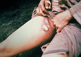
Sandfly BitesSigns and symptoms: The most common symptom of
cutaneous leishmaniasis is the development of skin sores or ulcers at the site of the sandfly bite.[158] They begin as nodules and can progress into volcanic ulcerative sores over a short period if untreated.
Visceral leishmaniasis human internal organs such as the liver, spleen, and bone marrow can develop within months and sometimes years after an infected sandfly bite.[159] Visceral leishmaniasis is a very serious illness and can result in death without treatment.
Diagnosis: Laboratory blood tests for leishmania antibodies can detect illness as well as tissue specimens from infected sores. Some tests are very difficult to perform and are only available at reference laboratories such as the Center for Disease Control and Prevention.[160]
Treatment: Cutaneous leishmaniasis should be treated with supportive and sanitary cleaning of the site to promote healing. Cutaneous leishmaniasis will heal on its own but careful monitoring is advised to prevent secondary infections. If cutaneous leishmaniasis does not heal it may progress into visceral infection and can be fatal.
Prevention: There are no current vaccines for leishmaniasis. Bite prevention is the best method of preventing infection in humans. Skin repellents can be used to deter sandfly bites as well as chemically treated clothing when exposed to endemic areas.[161]
References Information
Alkan, C., Bichaud, L., de Lamballerie, X., Alten, B., Gould, E. A., & Charrel, R. N. (2013). Sandfly-borne phleboviruses of eurasia and africa: Epidemiology, genetic diversity, geographic range, control measures. Antiviral Research, 100(1), 54-74. doi:10.1016/j.antiviral.2013.07.005 [doi]
Azad, A. F., Radulovic, S., Higgins, J. A., Noden, B. H., & Troyer, J. M. (1997). Flea-borne rickettsioses: Ecologic considerations. Emerging Infectious Diseases, 3(3), 319-327. doi:10.3201/eid0303.970308 [doi]
Beard, C.B., R.J. Eisen, C.M. Barker, J.F. Garofalo, M. Hahn, M. Hayden, A.J. Monaghan, N.H. Ogden, and P.J. Schramm, 2016: Ch. 5: Vectorborne Diseases. The Impacts of Climate Change on Human Health in the United States: A Scientific Assessment. U.S. Global Change Research Program, Washington, DC, 129–156. http://dx.doi.org/10.7930/J0765C7V
“Biology.” Centers for Disease Control and Prevention, Centers for Disease Control and Prevention, 1 Mar. 2016, www.cdc.gov/malaria/about/biology/index.html.
“Chikungunya.” World Health Organization, World Health Organization, www.who.int/denguecontrol/arbo-viral/other_arboviral_chikungunya/en/.
"Crimean-congo haemorrhagic fever." (2013). World Health Organization. Retrieved from http://www.who.int/mediacentre/factsheets/fs208/en/
“Dengue.” Centers for Disease Control and Prevention, Centers for Disease Control and Prevention, 3 Aug. 2017, www.cdc.gov/dengue/clinicallab/laboratory.html.
Dennis, D. T., & Chow, C. C. (2004). Plague. The Pediatric Infectious Disease Journal, 23(1), 69-71. doi:10.1097/01.inf.0000106918.18570.dd [doi]
Desjeux, P. (1992). Human leishmaniases: Epidemiology and public health aspects. World Health Statistics Quarterly.Rapport Trimestriel De Statistiques Sanitaires Mondiales, 45(2-3), 267-275.
Dionisio, D., Esperti, F., Vivarelli, A., & Valassina, M. (2003). Epidemiological, clinical and laboratory aspects of sandfly fever. Current Opinion in Infectious Diseases, 16(5), 383-388. doi:10.1097/01.qco.0000092808.64370.fa [doi]
Eisen, R. J., & Gage, K. L. (2012). Transmission of flea-borne zoonotic agents. Annual Review of Entomology, 57, 61-82. doi:10.1146/annurev-ento-120710-100717 [doi]
“Fact Sheet about Malaria.” World Health Organization, World Health Organization, www.who.int/mediacentre/factsheets/fs094/en/.
Flicek, B. F. (2007). Rickettsial and other tick-borne infections. Critical Care Nursing Clinics of North America, 19(1), 27-38. doi: S0899-5885(06)00122-5
Gulbudak, H., Cannataro, V. L., Tuncer, N., & Martcheva, M. (2017). Vector-borne pathogen and host evolution in a structured immuno-epidemiological system. Bulletin of Mathematical Biology, 79(2), 325-355. doi:10.1007/s11538-016-0239-0
Houseman, R. (2013). Guide to ticks and tick-borne diseases. Retrieved from http://extension.missouri.edu/publications/
Institute of Medicine (US) Forum on Microbial Threats. Vector-Borne Diseases: Understanding the Environmental, Human Health, and Ecological Connections, Workshop Summary. Washington (DC): National Academies Press (US); 2008. Summary and Assessment. Available from: https://www.ncbi.nlm.nih.gov/books/NBK52939/
“Japanese Encephalitis.” World Health Organization, World Health Organization, www.who.int/mediacentre/factsheets/fs386/en/.
Johnson, Arthur G. BRS Mircobiology and Immunology. Lippincott Williams & Wilkins, 2001.
Le, Tao, et al. First Aid for the USMLE Step 1. McGraw-Hill, 2015.
Leishmaniasis. (2013). Centers for Disease Control and Prevention. Retrieved from https://www.cdc.gov/parasites/leishmaniasis/
Lindquist, L., & Vapalahti, O. (2008). Tick-borne encephalitis. The Lancet, 371, December 10, 2017-1861-1871.53490-3.00011-X [doi]. Retrieved from
http://viala.org/img/TBE_seminar.pdf
"Lifecycle of blacklegged ticks." (2011). Centers for Disease Control and Prevention. Retrieved from https://www.cdc.gov/lyme/transmission/blacklegged.html
"Lyme disease. (2017)." Centers for Disease Control and Prevention. Retrieved from https://www.cdc.gov/lyme/
“Lymphatic Filariasis.” World Health Organization, World Health Organization, www.who.int/mediacentre/factsheets/fs102/en/.
Marinkelle, C. J. (1980). The control of leishmaniases. Bulletin of the World Health Organization, 58(6), 807–818.
Miller, E., Dushoff, J., & Huppert, A. (2016). The risk of incomplete personal protection coverage in vector-borne disease. Journal of the Royal Society Interface, 13(115), 20150666. http://doi.org.library1.unmc.edu:2048/10.1098/rsif.2015.0666
Pigott, D. M., Bhatt, S., Golding, N., Duda, K. A., Battle, K. E., Brady, O. J., … Hay, S. I. (2014). Global distribution maps of the leishmaniases. eLife, 3, e02851. http://doi.org.library1.unmc.edu:2048/10.7554/eLife.02851
"Rocky mountain spotted fever." (2017). Centers for Disease Control and Prevention. Retrieved from https://www.cdc.gov/rmsf/index.html
“Rift Valley Fever.” World Health Organization, World Health Organization, www.who.int/mediacentre/factsheets/fs207/en/.
Spach, D. H., Liles, W. C., Campbell, G. L., Quick, R. E., Anderson, D. E.,Jr, & Fritsche, T. R. (1993). Tick-borne diseases in the united states. The New England Journal of Medicine, 329(13), 936-947. doi:10.1056/NEJM199309233291308
Sun, Y.-C., Jarrett, C. O., Bosio, C. F., & Hinnebusch, B. J. (2014). Retracing the Evolutionary Path that Led to Flea-borne Transmission of Yersinia pestis. Cell Host & Microbe, 15(5), 578–586. http://doi.org.library1.unmc.edu:2048/10.1016/j.chom.2014.04.003
“The Human.” World Health Organization, World Health Organization, www.who.int/denguecontrol/human/en/.
"Tick-borne encephalitis." (2017). World Health Organization. Retrieved from http://www.who.int/ith/diseases/tbe/en/
"Tick-borne relapsing fever." (2015). Centers for Disease Control and Prevention. Retrieved from https://www.cdc.gov/relapsing-fever/index.html
"Tularemia." (2016)." Centers for Disease Control and Prevention. Retrieved from https://www.cdc.gov/tularemia/
Verwoerd, D. W. (2015). Definition of a vector and a vector-borne disease. Revue Scientifique Et Technique (International Office of Epizootics), 34(1), 29-39.
Wilson, A. J., Morgan, E. R., Booth, M., Norman, R., Perkins, S. E., Hauffe, H. C., … Fenton, A. (2017). What is a vector? Philosophical Transactions of the Royal Society B: Biological Sciences, 372(1719), 20160085. http://doi.org/10.1098/rstb.2016.0085
“West Nile Virus.” World Health Organization, World Health Organization, www.who.int/mediacentre/factsheets/fs354/en/.
Vector-borne diseases. World Health Organization, World Health Organization, Retrieved from http://www.who.int/mediacentre/factsheets/fs387/en/
“Zika Virus.” Centers for Disease Control and Prevention, Centers for Disease Control and Prevention, 21 June 2016, www.cdc.gov/zika/symptoms/index.html.
“Zika Virus.” World Health Organization, World Health Organization, www.who.int/mediacentre/factsheets/zika/en/.
^Prevention, CDC - Centers for Disease Control and.
"CDC - Leishmaniasis". www.cdc.gov. Retrieved 2017-12-15.
^Prevention, CDC - Centers for Disease Control and.
"CDC - Leishmaniasis". www.cdc.gov. Retrieved 2017-12-15.
^Prevention, CDC - Centers for Disease Control and.
"CDC - Leishmaniasis". www.cdc.gov. Retrieved 2017-12-15.
^Prevention, CDC - Centers for Disease Control and.
"CDC - Leishmaniasis". www.cdc.gov. Retrieved 2017-12-15.
^Prevention, CDC - Centers for Disease Control and.
"CDC - Leishmaniasis". www.cdc.gov. Retrieved 2017-12-15.
From Wikipedia, the free encyclopedia
Vector-borne diseases are human infections caused by
bacteria,
parasites, and
viruses that are spread by
vectors.[1] A vector is a living
organism that spreads
pathogens; in other words, it carries disease-causing organisms from one host to another. Vectors are normally considered to be
invertebrate animals, usually
arthropods, but also include
rodents, which carry the infectious disease from a reservoir to a susceptible host.[2] Vectors of human disease are typically a species of
mosquitoes,
ticks,
flies, and
fleas that are able to transmit bacteria, viruses, or parasites to humans and other vertebrate hosts.[3] A disease that is transmitted to humans, plants, or animals by any agent, arthropod, or fomite is a vector-borne disease.[4]
When trying to determine what the deadliest animal is in the world, a mosquito is likely not the first thing that comes to mind. However, the ability of mosquitoes to transmit disease causes millions of deaths each year. Female mosquitoes feed on the blood of infected animals and then spread the disease to humans through blood feeding. The diseases that they carry include malaria, dengue, yellow fever and countless others. There are treatments for some of these diseases, but for many there still is not treatment beyond supportive care. With the changing environmental climate, the concern about mosquito-borne disease becomes more serious with increases in flood zones and stagnant water. Preventative strategies include vaccinations when possible, wearing clothing to cover exposed areas, the use of insect repellent and destroying areas of breeding grounds.
Aedes Mosquito
Aedes Mosquito
Chikungunya
Vector type: Aedes aegypti and Aedes albopictus. This disease is found transmitted by these mosquito types in the Americas, Europe, Africa, Asia and India. When the breeding sites are close to homes they become a significant risk factor.[5]
Transmission: Chikungunya is transmitted via the bite of an infected female mosquito. These mosquitoes bite most frequently during the daylight, with peak activity occurring during the dawn and dusk. Recorded outbreaks are usually separated by period of 10 years.[6]
Signs and Symptoms: The onset of signs and symptoms of Chikungunya occur between 4-8 days and last for a duration of 2-3 days. The primary symptoms are fever with severe joint and muscle pain, headache, nausea, fatigue and rash. The name Chikyngunya means "to be contorted" because of the joint pain.[7] Most patients are able to fully recover but there are some serious complications that can occur. These complications involve the eyes, brain and heart and can cause death in older individuals. The signs and symptoms can often be confused with Dengue fever.
Diagnosis: Chikungunya is diagnosed with the use of serological tests. After infection, there is lifelong immunity against the disease.
Treatment: The most common ways to treat Chikungunya is with the use of supportive medicine. According to the WHO, this includes fluids, pain medicines and analgesics.[8] Unfortunately, there are currently no commercial vaccines available.
Prevention: The greatest preventative measures against the spread of Chikungunya involve mosquito protection. This can be done through the use of repellents, protective clothing, mosquito netting, window screens and breeding ground destruction.
Dengue Fever
Vector type: Aedes aegypti; There are 4 distinct serotypes of the Dengue virus that fall under that viral type of Flavivirus, Flaviviridae.[9]
Transmission: The disease is common in urban poor areas, suburbs and rural settings. The incidence has increased 30 times in the last 50 years, putting almost half of the worlds population at risk.[10]
Signs and Symptoms: Dengue presents with severe flu-like signs and symptoms and is at times referred to as "break-bone" fever. The onset of symptoms takes 4-10 days and lasts from 2-7 days. There is a severe headache, pain behind the eyes, nausea/vomiting, swollen glands, muscle and joint pains, rash, and skin lesions. Severe Dengue can also occur due to plasma leaking which causes an accumulation of fluid leading to respiratory distress and severe bleeding. This severe form of the disease is also known as dengue hemorrhagic fever. According to the WHO it and is one of the leading causes of death in Asian and Latin American countries. To detect the severe form of the disease patients should be monitored to see if after 3-7 days there is a drop in temperature. This is accompanied by severe abdominal pain, persistent vomiting, rapid breathing, bleeding gums, bloody vomit, fatigue and restlessness. A patient that recovers from the illness develops lifelong immunity from the specific serotype, but only partial from the other three infectious serotypes.[11]
Diagnosis: Dengue is detected via dengue NS1 serological testing.
Treatment: There is no specific treatment for Dengue. It is best to seek medical advice, rest and fluids. To treat the symptoms, paracetamol can be used to treat the fever and joint pains. However, it is important to avoid the use of aspirin or ibuprofen to decrease the bleeding risk.
Prevention: To prevent Dengue, it is important to follow mosquito protocol. It is also important to employ solid waste disposal and water storage practices.
Lymphatic Filariasis
also known as "elephantiasis"
Vector types: Culex in widespread urban and semi-urban areas. Anopheles in rural areas and Aedes in endemic islands in the Pacific.
Transmission: Lymphatic filiariasis is caused by an infection with nematodes (roundworms). The main types are Wuchereria bancrofti (90%) of cases, Brugia malaui and Brugia timori. Mosquitoes are infected with the microfilariae, which are then passed on to humans. The adults works then lodge into the lymphatic vessels and cause severe damage and lymphodema. This is usually acquired in childhood and leads to permanent disability.
Signs and Symptoms: There are three main divisions of the signs and symptoms associated with lymphatic filiariasis; asymptomatic, chronic and acute. In asymptomic infections, there are no external signs and symptoms of infection. However, there can still be some internal damage, especially to the lymphatics and kidneys. In chronic lymphatic filiariasis, there is lymphedema (tissue swelling), elephantiasis (skin/tissue thickening) of limbs and scrotal swelling (hydrocele). There can also be some involvement of the breasts and genitals.[12] The largest concern in those with chronic lymphatic filiariasis lies in the social stigma that follows. There is often severe stigma and physical disability that occurs as a results of this infection that perpetuates socioeconomic burdens, feelings of isolation and poverty. The last type of infection is acute lymphatic filiariasis. The acute infection has local inflammation of the skin, lymph nodes and lymphatic vessels. This can then lead to chronic lymphoedema or elephantiasis that can last for weeks. This occurs because of the immune response to the parasite and/or secondary bacterial skin infection due to the poor lymphatic drainage.
Treatment: The best treatment is through the use of preventative drug programs. This is done with a combined dose of albendazole+ivermetin or DEC to reduce the amount of microfiliariae.[13] By reducing the density of the parasite when it is small, the blockage of lymphatics by the adult worms can be prevented. It is important to note that this drug combination is ineffective against the adult forms. Other treatments include the relief of symptoms such as surgery for hydroceles.
Prevention: The WHO has implemented the Global Programme to Eliminate Lymphatic Filiariases (GPELF) in 2000.[14] The purpose of this program is to facilitate large scale annual treatments of preventative drug regimines in high risk areas. The program also includes increased management of those with disabilities resulting from infection with the end goal of decrease the stigma and socioeconomic burdens on these individuals. Mosquito protocol with insecticide treated nets, indoor spraying and personal protection measures are also encouraged.
Rift Valley Fever
Vector Type: Aedes and Culex mosquitoes.
Transmission: Rift Valley Fever occurs most commonly with direct/indirect contact with the blood or organs of infected animals. However, transmission can also occur if a mosquito is infected by feeding on an infected animal. Those who are most at risk include occupations like vets, herders, farmers and butchers. Transmission can also occur through inoculation in an open would or through inoculation.[15]
Signs and Symptoms: The onset of Rift Valley Fever symptoms occur between 2-6 days. Under normal conditions, it presents with flu-like signs and symptoms, fever, muscle and joint pain and headache. There have also been reports of neck stiffness, sensitivity to light, loss of appetite and vomiting. The signs and symptoms may be confused with those of meningitis. There is also a severe form of the disease that can take on three different forms. The first includes retinal lesions of the eyes. The second causes meninogoencephalitis with a presentation of intense headache, loss of memory, hallucinations, confusion, convulsions and even coma. These do not also appear immediately with onset of symptoms, but can occur more than 60 days post-infection. Lastly, there is a form associated with hemorrhagic fever. This occurs 2-4 days following the initial onset of symptoms. There is severe damage to the liver with jaundice/yellowing, vomiting blood, bleeding from the gums and nose and death between 3-6 days.[16]
Diagnosis: Rift Valley Fever can be diagnosed with RT-PCR assay, ELISA or viral isolation of cell culture
Treatment: There is no specified treatment, and medical intervention is supportive care.
Prevention: There is no commercially available vaccine, but there is an inactivated vaccine that can be used for at risk animals. It is best to closely monitor livestock in high risk areas to be on the lookout for outbreak.[17] It is also important to reduce exposure in at risk occupations and ensuring proper cooking of blood, meat and milk products. Lastly, mosquito protection is also important, especially in high risk seasons like following the rainy season.
Yellow Fever
Vector Type: Aedes mosquitoes (domestic, wild and semi-domestic forms)
Geography: endemic to Africa, Central and South America
Transmission: There are three types of transmission cycles associated with Yellow Fever; Jungle, Intermediate and Urban.[18] In the Jungle transmission cycle, the virus is transferred from monkeys to mosquitoes to humans. In the Intermediate cycle, the virus is spread by semi-domestic breeds of Aedes mosquitoes that are infected by monkeys and then infect humans through blood feeding. Due to increased contact between villages and areas of infection, there is increased transmission by this cycle, making is the most common type of outbreak in Africa. This also means that there can be outbreaks of disease in multiple villages occurring at the same time. Lastly, is the urban transmission cycle which is responsible for large epidemics in densely populated regions.[19]
Signs and Symptoms: Yellow Fever is an acute viral hemorrhagic disease. The associated symptoms unique to this disease include fever, headache, jaundice, muscle pain, nausea and vomiting, fatigue and black vomit. Most signs and symptoms only last 3-4 days. However, some may become severe cases which have a 50% mortality rate. The severe form occurs within the first 24 hours after it looks like recovery is taking place. The severe signs and symptoms present with a return of a high fever, severe jaundice with yellowing of the eyes and skin due to kidney and liver damage, dark urine and bleeding. This is a biphasic disease with the initial symptoms resulting from damage to the vascular endothelium, this then progresses to the liver during later replication.[18]
Diagnosis: Yellow Fever can be misdiagnosed as either severe malaria, viral hepatitis, poisoning or other types of hemorrhagic fevers when it is in the early phases. PCR and ELISA assays can be run to diagnose. A liver biopsy can also be done, you would expect to see Councilman bodies in the cytoplasm (esophinophilic hyaline masses).[19]
Treatment: There is no specific anti-viral drug therapy for infection with yellow fever. The recommended supportive treatment includes care for dehydration, fever and the complications from liver and kidney failure. Antibiotics may also be used to prevent any associated bacterial infections.
Prevention: There is a very successful vaccine that confers lifelong protection with a single dose and no need for a booster. The vaccine is an attenuated vaccine strain 17D, which is 99% effective within 30 days. [18]The vaccine is recommended in travelers to any endemic regions, in infants and even in the use of mass vaccination campaigns. It is also important to maintain mosquito protection protocol, added larvicides to water containers or any sites of stagnant water and routine insecticide spraying. Lastly, is maintaining epidemic preparedness. This results in fast detection with treatment and prevention response, which is crucial for controlling any outbreaks.
Zika
Vector Type: Aedes aegypti
Transmission: Zika is transmitted from the bite of an infected female mosquito when it feeds on humans. The most frequent times for infection occur during the day, especially at dawn and dusk when mosquitoes are the most prevalent. Sexual contact has also been found to be a pathway for transmission of Zika. [20]
Signs and Symptoms: Those infected present with a mild fever, skin rash, muscle and joint pain, headache or conjunctivitis that typically lasts for 2-7 days. The key features of Zika infection include the neurological components of microcephaly in pregnant women that is manifest in the fetus and Guilian-Barre syndrome with resulting transient paralysis.[21] Otherwise, it presents in a manner similar to that of the other arboviruses like Dengue.
Diagnosis: Zika can be detected through lab testing of blood, urine, saliva or semen.
Treatment: The recommended treatment options are largely supportive with rest, fluids and controlling pain and fever with normal drug regimens.
Prevention: There is no vaccine currently available for Zika. In order to protect against mosquito bites is most important with physical barriers like nets or covered clothing. Insect repellent that contains DEET, IR3535 or icaridin are other methods to prevent mosquitoes from feeding.[22] It is also important to detect and remove mosquito breeding sites. In terms of sexual transmission, safe sex practices and barriers like condoms are recommended, especially in women who are pregnant or may become pregnant.
Anopheles Mosquito
Anopheles Mosquito
Malaria
Vector Type: Anopheles species
Transmission: Malaria is transmitted via infection of a female mosquito with the plasmodium parasites; P. falciparum, P. vivax, P. ovale and P. malariae. According to the WHO, Africa carries the highest burden of disease with 90% of malaria cases and 91% of malarial deaths. Malaria infection is especially prevalent after the rainy season.[23] Those that are most at risk for becoming infected include infants, children, pregnant women and those with HIV/AIDS.
Signs and Symptoms: Malaria is an acute febrile illness that has an onset of symptoms that occur around 10-15 days after infection. The signs and symptoms include fever, headache and splenomegaly. The severity of disease is dependent on the type of plasmodium parasite.
P. Falciparum: The most severe form of the disease occurs with the plasmodium falciparum species that has a distinctive irregular fever pattern. In children, severe malaria can lead to severe anemia, respiratory distress as a result of metabolic acidosis and cerebral malaria. In adults, there can be multi-organ involvement and failure. The cause of these symptoms lies in the occlusion of capillaries by the parasitized red blood cells.[24]
P. vivax and P. ovale: These forms of disease are characterized by a fever pattern that has a 48 hour cycle with a fever occurring on the first and third days. There is a dormant form of these infections resulting from the hypnozoites in the liver.
P. malariae: This form of infection has a quartan fever pattern occuring every four days.
Diagnosis: Microscopy or rapid diagnostic testing can be done. P. falciparum can be diagnosed with the use of Giemsa staining with multiple ring forms and crescents seen. P. vivax and P. ovale can be diagnosed via Giemsa staining with red granules called Schuffner stippling throughout the red blood cell cytoplasm. P. malariae can be diagnosed through Giemsa staining with bar and band forms present.[19]
Treatment: The treatment of malaria can be complicated and depends on the type of infection and severity of disease. P. falciparum is treated with chloroquine and resistant strains are treated with quinine sulfate. P. vivas and ovale are treated with chloroquine and primaquine for the dormant liver form. Mefloquine or atovaquonone/proguanil can also be used to treat resistant strains. If the illness becomes life-threatening then IV quinidine or artesunate should be used, while making sure to test of G6PD deficiency.[19]
Prevention: Anti-malarial drugs and chemoprophylaxis are the most important in preventing the disease. Insecticide mosquito nets, indoor spraying with insecticides and disease surveillance are also important preventative measures. There is a vaccine available called Mosquirix that might provide some partial protection in young children.[24]
Lymphatic Filariasis
also known as "elephantiasis"
Vector types: Culex in widespread urban and semi-urban areas. Anopheles in rural areas and Aedes in endemic islands in the Pacific.
Transmission: Lymphatic filiariasis is caused by an infection with nematodes (roundworms). The main types are Wuchereria bancrofti (90%) of cases, Brugia malaui and Brugia timori. Mosquitoes are infected with the microfilariae, which are then passed on to humans. The adults works then lodge into the lymphatic vessels and cause severe damage and lymphodema. This is usually acquired in childhood and leads to permanent disability.
Signs and Symptoms: There are three main divisions of the signs and symptoms associated with lymphatic filiariasis; asymptomatic, chronic and acute. In asymptomic infections, there are no external signs and symptoms of infection. However, there can still be some internal damage, especially to the lymphatics and kidneys. In chronic lymphatic filiariasis, there is lymphedema (tissue swelling), elephantiasis (skin/tissue thickening) of limbs and scrotal swelling (hydrocele). There can also be some involvement of the breasts and genitals.[12] The largest concern in those with chronic lymphatic filiariasis lies in the social stigma that follows. There is often severe stigma and physical disability that occurs as a results of this infection that perpetuates socioeconomic burdens, feelings of isolation and poverty. The last type of infection is acute lymphatic filiariasis. The acute infection has local inflammation of the skin, lymph nodes and lymphatic vessels. This can then lead to chronic lymphoedema or elephantiasis that can last for weeks. This occurs because of the immune response to the parasite and/or secondary bacterial skin infection due to the poor lymphatic drainage.
Treatment: The best treatment is through the use of preventative drug programs. This is done with a combined dose of albendazole+ivermetin or DEC to reduce the amount of microfiliariae.[13] By reducing the density of the parasite when it is small, the blockage of lymphatics by the adult worms can be prevented. It is important to note that this drug combination is ineffective against the adult forms. Other treatments include the relief of symptoms such as surgery for hydroceles.
Prevention: The WHO has implemented the Global Programme to Eliminate Lymphatic Filiariases (GPELF) in 2000.[14] The purpose of this program is to facilitate large scale annual treatments of preventative drug regimines in high risk areas. The program also includes increased management of those with disabilities resulting from infection with the end goal of decrease the stigma and socioeconomic burdens on these individuals. Mosquito protocol with insecticide treated nets, indoor spraying and personal protection measures are also encouraged.
Culex Mosquito
Culex Mosquito
Japanese Encephalitis
Vector Type: Culex Species
Transmission: The transmission cycle includes mosquitoes, pigs, water birds and humans. Infection is particularly high in the warm, rainy and periods before the rice harvest. According to the WHO, Japanese encephalitis is the main cause of viral encephalitis in Asia with outbreaks occurring at intervals of 2-15 years.[25] Rural and semi-urban regions are the ones that are most prone to outbreak.
Signs and Symptoms: The most often types of signs and symptoms for Japanese encephalitis include a mild headache and fever. However, a severe form of the disease can occur. The presentation of the severe form of the disease includes a fast onset of high fever, headache, neck stiffness, disorientation, seizures, spastic paralysis and death. According to the WHO, the fatality rate can be as high as 30%. Of those that survive the severe form of JEV, 20-30% suffer permanent intellectual and neurological complications like paralysis, recurrent seizures and inability to speak.[26]
Diagnosis: lab testing of serum or cerebrospinal fluid is used if possible to obtain.
Treatment: There is no cure for Japanese Encephalitis. Intervention consists of relieving the clinical signs through supportive care.
Prevention: In addition to mosquito protocols, there are vaccines that can be used as parts of national immunization schedules. There are currently four types of vaccines available; inactivated mouse brain derived vaccines, inactivated Vero cell derived vaccines, live attenuated vaccines and live recombinant vaccines.[27]
Lymphatic Filariasis
also known as "elephantiasis"
Vector types: Culex in widespread urban and semi-urban areas. Anopheles in rural areas and Aedes in endemic islands in the Pacific.
Transmission: Lymphatic filiariasis is caused by an infection with nematodes (roundworms). The main types are Wuchereria bancrofti (90%) of cases, Brugia malaui and Brugia timori. Mosquitoes are infected with the microfilariae, which are then passed on to humans. The adults works then lodge into the lymphatic vessels and cause severe damage and lymphodema. This is usually acquired in childhood and leads to permanent disability.
Signs and Symptoms: There are three main divisions of the signs and symptoms associated with lymphatic filiariasis; asymptomatic, chronic and acute. In asymptomic infections, there are no external signs and symptoms of infection. However, there can still be some internal damage, especially to the lymphatics and kidneys. In chronic lymphatic filiariasis, there is lymphedema (tissue swelling), elephantiasis (skin/tissue thickening) of limbs and scrotal swelling (hydrocele). There can also be some involvement of the breasts and genitals.[12] The largest concern in those with chronic lymphatic filiariasis lies in the social stigma that follows. There is often severe stigma and physical disability that occurs as a results of this infection that perpetuates socioeconomic burdens, feelings of isolation and poverty. The last type of infection is acute lymphatic filiariasis. The acute infection has local inflammation of the skin, lymph nodes and lymphatic vessels. This can then lead to chronic lymphoedema or elephantiasis that can last for weeks. This occurs because of the immune response to the parasite and/or secondary bacterial skin infection due to the poor lymphatic drainage.
Treatment: The best treatment is through the use of preventative drug programs. This is done with a combined dose of albendazole+ivermetin or DEC to reduce the amount of microfiliariae.[13] By reducing the density of the parasite when it is small, the blockage of lymphatics by the adult worms can be prevented. It is important to note that this drug combination is ineffective against the adult forms. Other treatments include the relief of symptoms such as surgery for hydroceles.
Prevention: The WHO has implemented the Global Programme to Eliminate Lymphatic Filiariases (GPELF) in 2000.[14] The purpose of this program is to facilitate large scale annual treatments of preventative drug regimines in high risk areas. The program also includes increased management of those with disabilities resulting from infection with the end goal of decrease the stigma and socioeconomic burdens on these individuals. Mosquito protocol with insecticide treated nets, indoor spraying and personal protection measures are also encouraged.
West Nile Fever
Vector Type: Culex (species Pipiens)
Transmission: West Nile Fever is present in Africa, Europe, Middle East, North America and West Asia. The transmission cycle is between birds (crows and jay as natural hosts) and mosquitoes primarily, but also humans and horses.[28] There is also a very small percentage of infection from organ transplants, blood transfusions and breast milk.
Signs and Symptoms: 80% of West Nile infections are asymptomatic. However, for the other 20% there can be fevers, head and body aches, nausea, skin rash and swollen lymph glands. The incubation period before the onset of symptoms ranges from 3-14 days. There is also a severe form of the disease that has a neuroinvasive component. This presents with meningitis, high fever, neck stiffness, disorientation, convulsions, muscle weakness and paralysis. Those who are over 50 years old are most at risk. West Nile is the leading cause of arboviral encephalitis in the United States.[29]
Diagnosis: IgG antibody sero-conversion, ELISA-IgM, neutralization assays, PCR and viral isolation by cell culture is used to detect West Nile Fever.[19] IgM can also be detected in CSF and serum.
Treatment: Supportive care is recommended. For those with the neuroinvasive form of the disease, hospitalization with the use of IV fluids, respiratory support and antibiotics to prevent additional infection is recommended.[30]
Prevention: There are no vaccines for West Nile for humans, but ones that can be used for horses and to prevent transmission in horses.[31] An active animal health surveillance system can also be used to monitor outbreaks. Mosquito prevention is the most widely and effective use of prevention of infection.
Adult Deer Tick
Vector Type: Ticks Information
Ticks are the most significant vector of disease amongst wild and domestic animals in the world and are only second to mosquitoes in transmitting disease to humans.[32] In the United States, ticks are the leading cause of vector-borne infectious disease.[33]Ticks are external parasites that have been associated with the transmission of
pathogens for centuries dating back to the ancient times.[34] For ticks to complete their life-cycle, they are dependent on wild animals like deer and rodents for nourishment and transportation but also feed on domestic animals as hosts. There are two main families of ticks:
Ixodidae (
hard ticks) and
Argadidae (
soft ticks), that transmit disease to humans.[35] Hard ticks have a tough shield protecting their back and are darker in color while soft ticks do not have a protective shield, are lighter in color, and appear leathery.[36] Hard ticks present a substantial health risk and spread a majority of all
tick-borne infectious disease important to humans caused by bacteria, protozoa, and viruses. The life cycle of hard ticks is usually around two years during which they go through four growth phases: egg, larva, nymph, and adult.[37] In order for ticks to feed they must climb onto hosts (they do not jump or fly). One method to accomplish this is for ticks to wait for a host to pass by while sitting in a “questing” position characterized by grasping vegetation with their back legs and holding their front two legs out. When a host brushes by they quickly climb on and find a suitable place to feed.[38] Ticks can live in grasslands, wooded areas, wet or dry climates, and relatively temperate regions around the world in both North and South America, Africa, Europe, Asia, and Australia.
Crimean-Congo Hemorrhagic Fever (CCHF) is a widespread tick-borne disease caused by the transmission of Nairovirus which leads to viral hemorrhagic fever eruptions in humans. CCHF has a case-fatality rate of 10-40% and a mortality rate of 30%.[39] It is prevalent in the Middle East, Asia, the Balkins, Africa, and other countries south of the 50th parallel north.[40] Hosts of bunyaviridae ticks include domestic and wild animals such as cattle, sheep, goats, and ostriches in endemic areas.
Transmission: Crimean-Congo Hemorrhagic Fever (CCHF) is primarily transmitted to humans by tick bites or infected livestock animals.[41] Close contact with infected animal tissue immediately following the harvesting/slaughter of infected animals can cause infection in humans. Human-to-human transmission is possible with close contact to infected blood, secretions, organs, and bodily fluids. Hospital-acquired infections may happen due to unsanitary hygiene practices as well. Most CCHF infections occur in the livestock industry such as agricultural workers, veterinarians, and slaughterhouse workers.[42]
CCHF Skin LesionsSigns and symptoms: The incubation period for CCHF depends on how the Nairovirus was transmitted. Tick bite infections have an incubation period of 1-3 days (maximum of 9 days) before symptoms will show.[43] Infections caused by exposure to infected tissues or blood have an incubation period of 5-6 days (maximum of 13 days).[44] Symptoms of CCHF are sudden and may include fever, muscle aches, neck pain/stiffness, dizziness, headaches, sore eyes, backache, sensitivity to light, nausea, vomiting, sore throat, diarrhea, abdominal pain, and mood swings.[45] After 2-4 days symptoms may be replaced by sleepiness and depression and the abdominal pain may focus on the upper right abdominal area with detectable liver enlargement.[46] Clinical signs include fast heart rate, enlarged lymph nodes, and rashes in the mouth and throat as well as on the skin. Severely ill patients may progress into hepatitis and kidney, liver, and pulmonary failure after the fifth day. In severe cases, death usually occurs in the second week of infection.[47]
Diagnosis: CCHF can be diagnosed by a variety of laboratory tests including: enzyme-linked immunosorbent assay (ELISA), antigen detection, serum neutralization, reverse transcriptase polymerase chain reaction (RT-PCR) assay; and virus isolation by cell culture.[48]
Treatment: There is no absolute cure for CCHF. Treatment is achieved through supportive care for associated symptoms. An antiviral drug called ribavirin has be used to treat CCHF with notable benefit but lacks scientific evidence.[49]
Prevention: Controlling tick-borne infections like CCHF are difficult because of the level of difficulty in breaking the tick-animal-tick chain of infection (sequential infections; one after another). There are no current animal vaccinations and no clinically approved human vaccinations to prevent CCHF.[50] Raising awareness, using acaricides (chemicals to kill ticks), and personal protective equipment (long sleeves and pants, chemically treated clothing, skin repellent, light colored clothing to enhance visibility, gloves when handling livestock and tissues) are the best prevention methods to reduce infection.[51]
Lyme disease is the leading vector-borne disease in the United States with an estimated 329,000 cases diagnosed annually and one of the fastest growing vector-borne diseases in both the United States and Western Europe.[52] It was first identified in the United States in the mid-1970's during an investigation into an arthritis epidemic in Old Lyme, Connecticut.[53] Lyme Disease is caused by the bite of a deer tick which transmits the bacteria
borrelia burgdorferi. If untreated, Lyme disease can lead to debilitating illness affecting the joints, heart, brain, and nervous system.[54] Lyme disease predominately located in the Northeastern and upper Midwestern United States and Western Europe but can be found in the Pacific coast, Mid-Atlantic coast, and other parts of Europe.[55]
Transmission: Lyme Disease is spread by infected ixodes ticks when they bite hosts. In order for Lyme disease to be transmitted the tick must feed for a period of between 36-38 hours.[56] Ticks usually choose feeding areas in hard to detect places such as the groin, armpits, and scalp.[57] Most humans are infected by ticks in the nymph life stage due to their small size and difficulty to spot but, adult ticks may also infect humans as well.[58] Ixodes ticks feed on animals (most notably whitetail deer) and domestic pets like dogs and cats who then bring them into human domiciles where they can then climb onto humans. Wild and domestic animals can contract Lyme disease from infected ticks but there is no evidence that infected animals can transmit the disease to humans.[59] The incubation period for Lyme disease can range from anywhere between a few days up to a month (average time is 7 days) before the characteristic skin lesions appear.[60]
Erythema Migrans Skin LesionSigns and symptoms: The most notable sign of Lyme disease are skin lesions known as
“erythema migrans” (Latin for migrating redness) which occur in 90% of cases.[61][62] The skin lesions begin at the site of the bite and gradually expand up to a 12inch area.[63] Other associated symptoms are arthritis, headache, fatigue, fever, chills, muscle and joint soreness, and in later stages facial paralysis, heart palpitations, dizziness, shortness of breath, numbness and tingling in extremities, and memory loss.[64][65] End stages of the disease can lead to cranial neuritis, meningitis, and Bell’s palsy.
Diagnosis: Lyme disease can be diagnosed based on signs and symptoms with a history of exposure to endemic ixodes tick areas and laboratory tests for positive cultures of the borrelia burgdorferi bacteria.[66][67]
Treatment: Early stage treatment includes the administration of doxycycline, amoxicillin, cefuroxime, and azithromycin taken orally over a 2-3 week period.[68] Late stage treatment usually requires intravenous administration of penicillin or ceftriaxone over a 2-3 week period.[69]
Prevention: Reducing exposure to ticks is the best prevention method.[70] After exposure to tick endemic areas, thorough self-check procedures are recommended to reduce possibility of infection. Other preventive methods include wearing long sleeves and pants, chemically treat clothing with anti-tick chemicals, application of tick repellent to skin and probable feeding areas, and avoidance of contact with wildlife in endemic regions.
Tick-borne
Relapsing Fever (TBRF) is a bacterial infection that results in reoccurring (relapsing) fever, headache, muscle and joint pain, and nausea.[71] It is caused by
Borrelia bacteria which is transmitted through the bite of an
ornithodoros tick.[72] TBRF was first discovered in the United States in 1915.[73] The primary host of
ornithodoros ticks are rodents vice deer and domestic animals. Common hosts of these soft ticks are ground squirrels, chipmunks, prairie dogs, mice, and owls.[74] Soft ticks do not search for prey by “questing” in grasslands and wooded areas, rather they live in rodent burrows where they feed at will while rodents sleep and can survive and remain infectious for several years without feeding.[75][76] The average lifespan of soft ticks is around 10 years but can survive up to 20 years.[77] Most infections occur in the spring and summer months when humans vacation in mountainous rustic rodent infested cabins. TBRF is distributed throughout mountainous and plateau regions of North America, Mexico, South and Central America, the Mediterranean, Central Asia, and most of Africa.[78] In the United States, ornithodoros ticks are common in the western states of Texas, Arizona, California, Oklahoma, Colorado, Kansas, Montana, Idaho, Nevada, New Mexico, Oregon, Utah, and Wyoming.[79]
Transmission: TBRF is transmitted to humans through the bite of infected ornithodoros ticks. The bite is relatively painless and usually unnoticeable to humans. Soft ticks differ from hard ticks in that their bite only lasts for roughly 30-60 minutes during which time soft ticks feed on blood meal and transmit the
spirochete bacteria.[80] The incubation period of TBRF is on average 7 days but ranges from 4-18 days.[81]
Signs and symptoms: Primary symptoms of TBRF are high fever (around 103 degrees F), headache, muscle soreness, and joint pain. The signature characteristic of TBRF is that following incubation, onset of symptoms is sudden and persists for 3 days, followed by 7 days without symptoms, which is then followed by another 3-day period of symptoms.[82] This cycle can repeat several times (anywhere from 3 to 13 cycles) without treatment.[83] Death from TBRF is rare and usually only occurs in infants and elderly.[84]
Treatment: TBRF is treated with antibiotics such as penicillin, erythromycin, ceftriaxone, tetracyclines, macrolides, and fluoroquinolones.[86] An important note: when treating TBRF with antibiotics, patients should be observed for at 4 hours following administration.
Jarisch-Herxheimer reaction (a worsening of symptoms) occurs in close to 50% of all antibiotic treated patients.[87]
Prevention: Avoidance of exposure to rodent infestations, such as sleeping in mountain cabins with signs of rodent droppings and bedding, is recommended for prevention of TBRF.[88] Skin repellents (
DEET,
permethrin) can deter soft ticks from biting humans.
Rocky Mountain Spotted Fever (RMSF) is a microbial disease spread through tick bites and is caused by the pathogen Rickettsia ricketsii.[89] RMSF was first identified by
Howard Ricketts in the early 1900’s. Its name was derived from an outbreak of disease in the
Bitter Root Valley of western Montana.[90] Despite the name, RMSF can be found in Canada, Mexico, and throughout the United States and is prevalent in Southeastern states like North Carolina, Oklahoma, Tennessee, and South Carolina where 48% of U.S. cases are reported.[91] RMSF vectors are most active during the late spring and summer timeframes which is when greatest number of infections occur.[92] The most common vector of RMSF in the eastern states is the dog tick (Dermacentor variabilis) and the Rocky Mountain wood tick (Dermacentor andersoni) in the West.[93] The average incubation period for RMSF is 7 days but can range from 3 to 21days in some cases.[94]
Transmission: RMSF is transmitted through the bite of an infected tick. RMSF carrying hard ticks are usually transported to humans by domestic animals. These hard ticks will attach for 6-10 hours during which time Rickettsia ricketsii bacteria is transmitted to the human.[95]
RMSF Skin RashSigns and symptoms: Most of the early signs and symptoms of RMSF are not specific and are often overlooked as common cold symptoms. They include fever, nausea, headache, vomiting, stomach pain, muscle pain, and lack of appetite.[96] The most common sign of RMSF is the development of a rash that can very from pinpoint dots to red splotches and usually appear after 2-4 days after onset of symptoms.[97] Long term health effects of RMSF include amputation of affected extremities, hearing loss, paralysis, and mental disability.[98] In severe cases that go untreated, RMSF can result in death.
Diagnosis: Early diagnosis is difficult due to the generalized symptoms. Diagnosis can be confirmed with the development of rashes and blood tests.
Treatment: The recommended treatment is doxycycline for both adults and children of all ages.[99]
Prevention: There is no vaccine for RMSF. Prevention is primarily achieved through preventing tick bites and preventing ticks from attaching to domestic animals.[100] Reducing exposure to ticks is the best prevention method. After exposure to tick endemic areas, through self-check and animal check procedures are recommended to reduce possibility of infection. Other preventive methods include wearing long sleeves and pants, chemically treat clothing with anti-tick chemicals, application of tick repellent (
DEET,
permethrin,
picaridin) to skin and probable feeding areas, and avoidance of contact with wildlife in endemic regions.
Tick-borne Encephalitis (TBE) is a viral infectious disease affecting the human
central nervous system.[101] It is caused by the transmission of the Flaviviridae virus via ixodes ticks.[102] TBE was first clinically discovered in 1931 in Austria and later isolated as a causative agent in Eastern Russia in 1937.[103] The primary hosts for these hard ticks are rodents while other large animals act as feeding hosts as well but do not play a role in the maintenance of the virus. TBE is endemic to Russia, the Baltic States and Slovenia and can be found in Albania, Austria, Belarus, Bosnia, Bulgaria, China, Croatia, Denmark, Finland, Germany, Greece, Hungary, Italy, Mongolia, Norway, Poland, the Republic of Korea, Romania, Serbia, Slovakia, Sweden, Switzerland, Turkey and Ukraine. TBE carrying ixodes tick activity usually begins in the spring and lasts through November.[104]
Transmission: TBE is mainly transmitted via the bite of an infected ixodes tick. Infection may also be transmitted through the consumption of raw milk from infected agricultural animals such as cows, goats, and sheep.[105] Infected ixodes ticks will attach to a host for several days but the transmission of the Flaviviridae virus happens within minutes, so early removal will not prevent transmission.[106] Incubation period for TBE ranges between 7-14 days (shorter periods have been reported).[107]
Signs and symptoms: Early symptoms of infection include fever, malaise, anorexia, muscle aches, headache, nausea, and vomiting.[108] After a short remission period (7-8 days) a second stage of symptoms affecting the central nervous system present.[109] These include signs of meningitis (fever, headache, and a stiff neck) and encephalitis (drowsiness, confusion, sensory disturbances, and paralysis).[110] Although rare, TBE can result in death if untreated.
Diagnosis: Clinical diagnosis during the first stage depends the detection of the virus by
RT-PCR from blood testing.[111] TBE is not usually detected in the first stage of infection but, TBE
immunoglobulin and TBE antibodies are present in blood samples taken in the second stage of symptoms and can be identified.[112]
Treatment: There is no sure for TBE. Treatment is achieved through supportive care of symptoms.
Prevention: As with other tick-borne diseases, prevention relies on preventing ticks from biting animals and humans. There is a vaccine available for endemic areas however, adverse reactions to the vaccine in children limit its use.[113]
Tularemia is highly infectious ulcerative disease caused by the coccobacilli bacteria
Francisella tularensis which can be transmitted by the bite of an ixodes
dog tick,
wood tick and
lone star tick.[114][115] The causative agent was first isolated from infected rodents during the early 1900’s in
Tulare County, California.[116] The most important tick-borne hosts of tularemia are rabbits and hares but are also found on domestic cats and hamsters, muskrats, prairie dogs, and other rodents.[117] Tularemia vectors can be found in Southeastern, South-Central, and Western United States and are prevalent in Arkansas, Oklahoma, and Missouri. [118]
Transmission: Infection usually occurs via the bite of an infected tick but can also be transmitted through skin contact with infected animal tissues (hunting, skinning), and the consumption of infected animal tissue.[119] The incubation period for tick-borne tularemia range from 1-14 days with an average of 3-5 days before onset of symptoms.[120]
Signs and symptoms: Tick-borne tularemia initially presents with fever, chills, headache, body ache, and fatigue.[121] A classic sign of tick-borne tularemia is the development of a skin ulcer at the site of the bite accompanied by glandular swelling usually in the groin or armpit.[122] Depending on the severity of the infection, symptoms can progress into secondary pneumonia, mild hepatitis, and pharyngitis.[123]
Diagnosis: Tularemia can be difficult to diagnose due to its rarity and the commonality of its associated symptoms. Patient disclosure of possible tick bite or exposure to wild animals is important to diagnosis.[124] Blood tests and cultures can be used to confirm diagnosis.[125]
Treatment : Administration of antibiotics like
streptomycin,
gentamicin,
doxycycline, or
ciprofloxacin over a 10-21 day period is the preferred method of treatment.[126] Despite symptoms persisting several weeks, with antibiotic treatment, most infected humans have a full recovery.
Prevention: As with other tick-borne diseases, prevention relies on preventing tick bites. Reducing exposure to ticks is the best prevention method. After exposure to tick endemic areas, through self-check procedures are recommended to reduce possibility of infection. Other preventive methods include wearing long sleeves and pants, using gloves to handle dead animals and during skinning procedures, chemically treat clothing with anti-tick chemicals, application of tick repellent to skin and probable feeding areas, and avoidance of contact with wildlife in endemic regions.
Vector Type: Fleas Information
Fleas have historically presented a substantial health risk to the human population as they were the source of plague pandemics like the Black Death and the Great Plague in the 1330’s and 1660’s and claimed over 50 million lives. Fleas are geographically disbursed worldwide and can be found on all inhabited continents. Rapidly changing ecological and environmental factors have allowed for flea-borne diseases of the past to reemerge. In residential areas, vector fleas are most often introduced to the human populous through their natural hosts: rodents and domestic animals. Fleas commonly found on free-ranging cats and dogs, opossums, raccoons, and squirrels are picked up by household pets and brought into homes. Aside from being a nuisance, these fleas may carry disease causing organisms. Of the 2,000 species and subspecies of fleas, only a handful serve as vectors of human diseases. There are several disease causing bacteria of public health importance that are maintained and transmitted by fleas, among them, Yersinia pestis and Rickettsia typhi the causative agents of plague and murine typhus (rickettsiosis). Due to the frequent feeding behavior of fleas and their extraordinary mobility, pathogens spread rapidly to human populations. Fleas have a four-stage life cycle involving eggs, larvae, pupae, and adults. Eggs are laid by the female in the environment and hatch into larvae after a 3-4 day period and feed on organic debris in the environment. Larvae eventually form cocoon or pupae protected by debris from the environment. It usually takes about 3-4 weeks for the larval and pupal stages to complete. Afterwards, adults hatch from pupae and seek out a warm-blooded host for blood meals.
The plague is caused by a bacterium known as
Yersinia pestis and is transmitted to humans through bites from infected fleas.[127] Diamanus montanus and
Pulex irritans are two of the main plague vectors that transmit the disease to humans. Plague bacteria maintain existence through an
enzootic cycle being passed from infected rodents to fleas and back to other rodents until contact is made with either humans or domestic animals in which case the bacteria are spread.[128] Wild hosts of plague vector fleas include rock squirrels, mice, wood rats, prairie dogs, chipmunks, ground squirrels, voles, and rabbits.[129] Carnivorous animals can also become infected by eating infected rodents.
Bubonic Plague BuboesTransmission: The primary route of transmission for plague to humans is through infected flea bites. Plague outbreaks usually occur during
epizootics when several infected rodents die and hungry fleas seek out new hosts.[130] Humans and domestic animals that visit areas where these rodents die are at high risk for disease transmission.[131] Other modes of transmission are through skin contact with infected animal tissues (hunting, skinning, rodent clean-up) and by inhaling aerosolized droplets from humans infected with pneumonic plague.
Signs and symptoms: The three most common clinical forms of plague are
bubonic,
pneumonic, and
septicemic.[132] Bubonic plague has an incubation period of 1-7 days (typically 3-5).[133] Early symptoms include fever, headache, chills and weakness. Within 24 hours of symptoms, a lymph node (
bubo) close to the site of the bite will swell and in 25% of cases a skin ulcer will form at the site.[134] Without treatment, vomiting, anorexia, abdominal pain, and altered mental status may develop and lead to septicemic plague. Septicemic plague can cause multi-organ system failure including target organs like heart, kidney, spleen, lung, liver, skin, and the central nervous system.[135] Pneumonic plague can result from untreated septicemic and bubonic plague characterized by the rapid development of pneumonia and can lead to respiratory failure and shock.[136] Untreated forms of plague can result in death with pneumonic fatality occurring within 24 hours.[137]
Diagnosis: Plague is most readily diagnosed by the identification of a
bubo and disclosure of recent exposure to endemic areas or flea bites. Blood and bubo samples can be testes by through laboratory procedures to confirm diagnosis.[138]
Treatment: Although plague is a serious illness, it can be treated with common antibiotics including streptomycin, gentamycin, tetracycline, and doxycycline.[139]
Prevention: Vaccination is only recommended for highly exposed personnel working with plague specimens. Preventive measures include educating and raising awareness for people when zoonotic plague is present in their area and advising them to take precautions against flea bites and not to handle animal carcasses. Avoidance of contact with bodily infected fluids and tissues is generally advised.[140]
Flea-borne
rickettsiosis (also called,
murine typhus,
endemic typhus, and flea-borne typhus) is a disease cause by the infection of bacteria known as Rickettsia typhi and is primarily transmitted through contact with infected Xenopsylla cheopis fleas.[141] Murine typhus is widely distributed along oceanic coastal regions in tropical and sub-tropical areas throughout the world.[142] Rats are the primary host of murine typhus carrying fleas but they can also be found on cats and opossums.[143]
Transmission: The Rickettsia typhi bacteria is transmitted to humans when contact with infected fleas is made. Murine typhus carrying fleas infect humans when infected flea feces is rubbed into cuts, scrapes, or at bites sites.[144]
Signs and symptoms: The incubation period for flea-borne rickettsiosis is around 14 days after which onset of fever and chills, body ache, appetite loss, nausea, vomiting, stomach pain, and cough usually occur.[145] After about 5 days, a rash may start to form around the area of infection. Untreated infections can progress into severe complication including damage to liver, kidneys, lungs, heart, and brain.[146]
Diagnosis: Murine typhus diagnosis is difficult due to its symptoms closely relating to many other illnesses and is dependent on disclosure of recent exposure to endemic areas or flea bites. Blood tests can confirm diagnosis but may take several weeks to complete.[147]
Treatment: The most common treatment of Murine typhus is with the antibiotic doxycycline which can be administered to all ages.[148]
Prevention: There is no current vaccine for murine typhus. Reducing exposure to fleas and flea infested areas is the best prevention method. Other preventive methods include wearing long sleeves and pants, using gloves to handle dead animals, chemically treat clothing with DEET, application of flea repellent to skin, and avoidance of contact with wildlife in endemic regions.[149]
Vector Type: Sandflies Information
Phlebotomine sandflies are widely distributed throughout the world occurring in Africa, Europe, the Middle East, and Asia.[150] Warm, moist, and humid climates provide the best environments for sandflies to thrive in as the moisture is needed for egg survival. If temperatures drop below 10 degrees Celsius, sandflies must enter a dormant period in order to survive to winter.[151] Sandflies have elongated piercing mouthparts for taking blood and are generally a third of the size of a mosquito. The life cycle of the sandfly has 4 stages: egg, larva, pupal, and adult which can take anywhere from 26-43 days depending on environmental factors.[152]
Leishmaniasis is a parasitic infection spread via the bite of an infected female
phlebotominesandfly. Leishmania parasites can be found in over 88 countries throughout the world but are endemic to Africa, South and Central America, Asia, and southern Europe.[153] Although widely distributed, 90% of potentially fatal cases are reported in 6 only countries: Brazil, India, Ethiopia, Sudan, South Sudan, and Bangladesh.[154] Leishmania reservoir hosts are dogs, foxes, jackals, sloths, rodents, marsupials, primates and other mammals.[155] Phlebotomine sandflies become infected after taking blood meal from any of these infected hosts and then can transmit the disease to humans.[156]
Transmission: Leishmania parasites are transmitted to humans when the infected sandfly pierces the skin to take blood meal.[157]
Sandfly BitesSigns and symptoms: The most common symptom of
cutaneous leishmaniasis is the development of skin sores or ulcers at the site of the sandfly bite.[158] They begin as nodules and can progress into volcanic ulcerative sores over a short period if untreated.
Visceral leishmaniasis human internal organs such as the liver, spleen, and bone marrow can develop within months and sometimes years after an infected sandfly bite.[159] Visceral leishmaniasis is a very serious illness and can result in death without treatment.
Diagnosis: Laboratory blood tests for leishmania antibodies can detect illness as well as tissue specimens from infected sores. Some tests are very difficult to perform and are only available at reference laboratories such as the Center for Disease Control and Prevention.[160]
Treatment: Cutaneous leishmaniasis should be treated with supportive and sanitary cleaning of the site to promote healing. Cutaneous leishmaniasis will heal on its own but careful monitoring is advised to prevent secondary infections. If cutaneous leishmaniasis does not heal it may progress into visceral infection and can be fatal.
Prevention: There are no current vaccines for leishmaniasis. Bite prevention is the best method of preventing infection in humans. Skin repellents can be used to deter sandfly bites as well as chemically treated clothing when exposed to endemic areas.[161]
References Information
Alkan, C., Bichaud, L., de Lamballerie, X., Alten, B., Gould, E. A., & Charrel, R. N. (2013). Sandfly-borne phleboviruses of eurasia and africa: Epidemiology, genetic diversity, geographic range, control measures. Antiviral Research, 100(1), 54-74. doi:10.1016/j.antiviral.2013.07.005 [doi]
Azad, A. F., Radulovic, S., Higgins, J. A., Noden, B. H., & Troyer, J. M. (1997). Flea-borne rickettsioses: Ecologic considerations. Emerging Infectious Diseases, 3(3), 319-327. doi:10.3201/eid0303.970308 [doi]
Beard, C.B., R.J. Eisen, C.M. Barker, J.F. Garofalo, M. Hahn, M. Hayden, A.J. Monaghan, N.H. Ogden, and P.J. Schramm, 2016: Ch. 5: Vectorborne Diseases. The Impacts of Climate Change on Human Health in the United States: A Scientific Assessment. U.S. Global Change Research Program, Washington, DC, 129–156. http://dx.doi.org/10.7930/J0765C7V
“Biology.” Centers for Disease Control and Prevention, Centers for Disease Control and Prevention, 1 Mar. 2016, www.cdc.gov/malaria/about/biology/index.html.
“Chikungunya.” World Health Organization, World Health Organization, www.who.int/denguecontrol/arbo-viral/other_arboviral_chikungunya/en/.
"Crimean-congo haemorrhagic fever." (2013). World Health Organization. Retrieved from http://www.who.int/mediacentre/factsheets/fs208/en/
“Dengue.” Centers for Disease Control and Prevention, Centers for Disease Control and Prevention, 3 Aug. 2017, www.cdc.gov/dengue/clinicallab/laboratory.html.
Dennis, D. T., & Chow, C. C. (2004). Plague. The Pediatric Infectious Disease Journal, 23(1), 69-71. doi:10.1097/01.inf.0000106918.18570.dd [doi]
Desjeux, P. (1992). Human leishmaniases: Epidemiology and public health aspects. World Health Statistics Quarterly.Rapport Trimestriel De Statistiques Sanitaires Mondiales, 45(2-3), 267-275.
Dionisio, D., Esperti, F., Vivarelli, A., & Valassina, M. (2003). Epidemiological, clinical and laboratory aspects of sandfly fever. Current Opinion in Infectious Diseases, 16(5), 383-388. doi:10.1097/01.qco.0000092808.64370.fa [doi]
Eisen, R. J., & Gage, K. L. (2012). Transmission of flea-borne zoonotic agents. Annual Review of Entomology, 57, 61-82. doi:10.1146/annurev-ento-120710-100717 [doi]
“Fact Sheet about Malaria.” World Health Organization, World Health Organization, www.who.int/mediacentre/factsheets/fs094/en/.
Flicek, B. F. (2007). Rickettsial and other tick-borne infections. Critical Care Nursing Clinics of North America, 19(1), 27-38. doi: S0899-5885(06)00122-5
Gulbudak, H., Cannataro, V. L., Tuncer, N., & Martcheva, M. (2017). Vector-borne pathogen and host evolution in a structured immuno-epidemiological system. Bulletin of Mathematical Biology, 79(2), 325-355. doi:10.1007/s11538-016-0239-0
Houseman, R. (2013). Guide to ticks and tick-borne diseases. Retrieved from http://extension.missouri.edu/publications/
Institute of Medicine (US) Forum on Microbial Threats. Vector-Borne Diseases: Understanding the Environmental, Human Health, and Ecological Connections, Workshop Summary. Washington (DC): National Academies Press (US); 2008. Summary and Assessment. Available from: https://www.ncbi.nlm.nih.gov/books/NBK52939/
“Japanese Encephalitis.” World Health Organization, World Health Organization, www.who.int/mediacentre/factsheets/fs386/en/.
Johnson, Arthur G. BRS Mircobiology and Immunology. Lippincott Williams & Wilkins, 2001.
Le, Tao, et al. First Aid for the USMLE Step 1. McGraw-Hill, 2015.
Leishmaniasis. (2013). Centers for Disease Control and Prevention. Retrieved from https://www.cdc.gov/parasites/leishmaniasis/
Lindquist, L., & Vapalahti, O. (2008). Tick-borne encephalitis. The Lancet, 371, December 10, 2017-1861-1871.53490-3.00011-X [doi]. Retrieved from
http://viala.org/img/TBE_seminar.pdf
"Lifecycle of blacklegged ticks." (2011). Centers for Disease Control and Prevention. Retrieved from https://www.cdc.gov/lyme/transmission/blacklegged.html
"Lyme disease. (2017)." Centers for Disease Control and Prevention. Retrieved from https://www.cdc.gov/lyme/
“Lymphatic Filariasis.” World Health Organization, World Health Organization, www.who.int/mediacentre/factsheets/fs102/en/.
Marinkelle, C. J. (1980). The control of leishmaniases. Bulletin of the World Health Organization, 58(6), 807–818.
Miller, E., Dushoff, J., & Huppert, A. (2016). The risk of incomplete personal protection coverage in vector-borne disease. Journal of the Royal Society Interface, 13(115), 20150666. http://doi.org.library1.unmc.edu:2048/10.1098/rsif.2015.0666
Pigott, D. M., Bhatt, S., Golding, N., Duda, K. A., Battle, K. E., Brady, O. J., … Hay, S. I. (2014). Global distribution maps of the leishmaniases. eLife, 3, e02851. http://doi.org.library1.unmc.edu:2048/10.7554/eLife.02851
"Rocky mountain spotted fever." (2017). Centers for Disease Control and Prevention. Retrieved from https://www.cdc.gov/rmsf/index.html
“Rift Valley Fever.” World Health Organization, World Health Organization, www.who.int/mediacentre/factsheets/fs207/en/.
Spach, D. H., Liles, W. C., Campbell, G. L., Quick, R. E., Anderson, D. E.,Jr, & Fritsche, T. R. (1993). Tick-borne diseases in the united states. The New England Journal of Medicine, 329(13), 936-947. doi:10.1056/NEJM199309233291308
Sun, Y.-C., Jarrett, C. O., Bosio, C. F., & Hinnebusch, B. J. (2014). Retracing the Evolutionary Path that Led to Flea-borne Transmission of Yersinia pestis. Cell Host & Microbe, 15(5), 578–586. http://doi.org.library1.unmc.edu:2048/10.1016/j.chom.2014.04.003
“The Human.” World Health Organization, World Health Organization, www.who.int/denguecontrol/human/en/.
"Tick-borne encephalitis." (2017). World Health Organization. Retrieved from http://www.who.int/ith/diseases/tbe/en/
"Tick-borne relapsing fever." (2015). Centers for Disease Control and Prevention. Retrieved from https://www.cdc.gov/relapsing-fever/index.html
"Tularemia." (2016)." Centers for Disease Control and Prevention. Retrieved from https://www.cdc.gov/tularemia/
Verwoerd, D. W. (2015). Definition of a vector and a vector-borne disease. Revue Scientifique Et Technique (International Office of Epizootics), 34(1), 29-39.
Wilson, A. J., Morgan, E. R., Booth, M., Norman, R., Perkins, S. E., Hauffe, H. C., … Fenton, A. (2017). What is a vector? Philosophical Transactions of the Royal Society B: Biological Sciences, 372(1719), 20160085. http://doi.org/10.1098/rstb.2016.0085
“West Nile Virus.” World Health Organization, World Health Organization, www.who.int/mediacentre/factsheets/fs354/en/.
Vector-borne diseases. World Health Organization, World Health Organization, Retrieved from http://www.who.int/mediacentre/factsheets/fs387/en/
“Zika Virus.” Centers for Disease Control and Prevention, Centers for Disease Control and Prevention, 21 June 2016, www.cdc.gov/zika/symptoms/index.html.
“Zika Virus.” World Health Organization, World Health Organization, www.who.int/mediacentre/factsheets/zika/en/.


.jpg)


.jpg)