Single-strand DNA-binding protein (SSB) is a protein found in Escherichia coli (E. coli) bacteria, that binds to single-stranded regions of deoxyribonucleic acid ( DNA). [1] Single-stranded DNA is produced during all aspects of DNA metabolism: replication, recombination, and repair. As well as stabilizing this single-stranded DNA, SSB proteins bind to and modulate the function of numerous proteins involved in all of these processes.
Active E. coli SSB is composed of four identical 19 kDa subunits. Binding of single-stranded DNA to the tetramer can occur in different "modes", with SSB occupying different numbers of DNA bases depending on a number of factors, including salt concentration. For example, the (SSB)65 binding mode, in which approximately 65 nucleotides of DNA wrap around the SSB tetramer and contact all four of its subunits, is favoured at high salt concentrations in vitro. At lower salt concentrations, the (SSB)35 binding mode, in which about 35 nucleotides bind to only two of the SSB subunits, tends to form. Further work is required to elucidate the functions of the various binding modes in vivo.
Bacterial SSB
| SSB | |||||||||
|---|---|---|---|---|---|---|---|---|---|
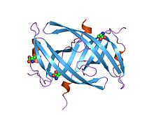
 Crystal structure of PriB- a primosomal DNA replication protein of Escherichia coli | |||||||||
| Identifiers | |||||||||
| Symbol | SSB | ||||||||
| Pfam | PF00436 | ||||||||
| Pfam clan | CL0021 | ||||||||
| InterPro | IPR000424 | ||||||||
| PROSITE | PDOC00602 | ||||||||
| SCOP2 | 1kaw / SCOPe / SUPFAM | ||||||||
| TCDB | 3.A.7 | ||||||||
| |||||||||
SSB protein domains in bacteria are important in its function of maintaining DNA metabolism, more specifically DNA replication, repair, and recombination. [2] It has a structure of three beta-strands to a single six-stranded beta-sheet to form a protein dimer. [3]
See also
References
- ^ Shishmarev D, Wang Y, Mason CE, Su XC, Oakley AJ, Graham B, et al. (February 2014). "Intramolecular binding mode of the C-terminus of Escherichia coli single-stranded DNA binding protein determined by nuclear magnetic resonance spectroscopy". Nucleic Acids Research. 42 (4): 2750–2757. doi: 10.1093/nar/gkt1238. PMC 3936761. PMID 24288378.
- ^ Meyer RR, Laine PS (December 1990). "The single-stranded DNA-binding protein of Escherichia coli". Microbiological Reviews. 54 (4): 342–380. doi: 10.1128/MMBR.54.4.342-380.1990. PMC 372786. PMID 2087220.
- ^ Raghunathan S, Ricard CS, Lohman TM, Waksman G (June 1997). "Crystal structure of the homo-tetrameric DNA binding domain of Escherichia coli single-stranded DNA-binding protein determined by multiwavelength x-ray diffraction on the selenomethionyl protein at 2.9-A resolution". Proceedings of the National Academy of Sciences of the United States of America. 94 (13): 6652–6657. doi: 10.1073/pnas.94.13.6652. PMC 21213. PMID 9192620.
External links
- Single-Stranded+DNA+Binding+Proteins at the U.S. National Library of Medicine Medical Subject Headings (MeSH)
- SSB in PFAM
Single-strand DNA-binding protein (SSB) is a protein found in Escherichia coli (E. coli) bacteria, that binds to single-stranded regions of deoxyribonucleic acid ( DNA). [1] Single-stranded DNA is produced during all aspects of DNA metabolism: replication, recombination, and repair. As well as stabilizing this single-stranded DNA, SSB proteins bind to and modulate the function of numerous proteins involved in all of these processes.
Active E. coli SSB is composed of four identical 19 kDa subunits. Binding of single-stranded DNA to the tetramer can occur in different "modes", with SSB occupying different numbers of DNA bases depending on a number of factors, including salt concentration. For example, the (SSB)65 binding mode, in which approximately 65 nucleotides of DNA wrap around the SSB tetramer and contact all four of its subunits, is favoured at high salt concentrations in vitro. At lower salt concentrations, the (SSB)35 binding mode, in which about 35 nucleotides bind to only two of the SSB subunits, tends to form. Further work is required to elucidate the functions of the various binding modes in vivo.
Bacterial SSB
| SSB | |||||||||
|---|---|---|---|---|---|---|---|---|---|
 Crystal structure of PriB- a primosomal DNA replication protein of Escherichia coli | |||||||||
| Identifiers | |||||||||
| Symbol | SSB | ||||||||
| Pfam | PF00436 | ||||||||
| Pfam clan | CL0021 | ||||||||
| InterPro | IPR000424 | ||||||||
| PROSITE | PDOC00602 | ||||||||
| SCOP2 | 1kaw / SCOPe / SUPFAM | ||||||||
| TCDB | 3.A.7 | ||||||||
| |||||||||
SSB protein domains in bacteria are important in its function of maintaining DNA metabolism, more specifically DNA replication, repair, and recombination. [2] It has a structure of three beta-strands to a single six-stranded beta-sheet to form a protein dimer. [3]
See also
References
- ^ Shishmarev D, Wang Y, Mason CE, Su XC, Oakley AJ, Graham B, et al. (February 2014). "Intramolecular binding mode of the C-terminus of Escherichia coli single-stranded DNA binding protein determined by nuclear magnetic resonance spectroscopy". Nucleic Acids Research. 42 (4): 2750–2757. doi: 10.1093/nar/gkt1238. PMC 3936761. PMID 24288378.
- ^ Meyer RR, Laine PS (December 1990). "The single-stranded DNA-binding protein of Escherichia coli". Microbiological Reviews. 54 (4): 342–380. doi: 10.1128/MMBR.54.4.342-380.1990. PMC 372786. PMID 2087220.
- ^ Raghunathan S, Ricard CS, Lohman TM, Waksman G (June 1997). "Crystal structure of the homo-tetrameric DNA binding domain of Escherichia coli single-stranded DNA-binding protein determined by multiwavelength x-ray diffraction on the selenomethionyl protein at 2.9-A resolution". Proceedings of the National Academy of Sciences of the United States of America. 94 (13): 6652–6657. doi: 10.1073/pnas.94.13.6652. PMC 21213. PMID 9192620.
External links
- Single-Stranded+DNA+Binding+Proteins at the U.S. National Library of Medicine Medical Subject Headings (MeSH)
- SSB in PFAM