| S4 | |||||||||
|---|---|---|---|---|---|---|---|---|---|
 | |||||||||
| Identifiers | |||||||||
| Symbol | S4 | ||||||||
| Pfam | PF01479 | ||||||||
| Pfam clan | CL0492 | ||||||||
| InterPro | IPR002942 | ||||||||
| PROSITE | PDOC00549 | ||||||||
| SCOP2 | 1c06 / SCOPe / SUPFAM | ||||||||
| CDD | cd00165 | ||||||||
| |||||||||
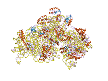
In molecular biology, S4 domain refers to a small RNA-binding protein domain found in a ribosomal protein named uS4 (called S9 in eukaryotes). The S4 domain is approximately 60-65 amino acid residues long, occurs in a single copy at various positions in different proteins and was originally found in pseudouridine syntheses, a bacterial ribosome-associated protein. [1]
The S4 protein helps to initiate assembly of the 16S rRNA. In this way proteins serve to organise and stabilise the rRNA tertiary structure. [2] [3]
Function
The function of the S4 domain is to be an RNA-binding protein. S4 is a multifunctional protein, and it must bind to the 16S ribosomal RNA. In addition, the S4 domain binds a complex pseudoknot and represses translation. More specifically, this protein domain delivers nucleotide-modifying enzymes to RNA and to regulates translation through structure specific RNA binding. [1]
Structure
The S4 protein domain is composed of three alpha helices and five beta strands. It is organized as an antiparallel sheet in a Greek key motif. [4]
References
- ^ a b Aravind L, Koonin EV (March 1999). "Novel predicted RNA-binding domains associated with the translation machinery". J. Mol. Evol. 48 (3): 291–302. doi: 10.1007/pl00006472. PMID 10093218. S2CID 8083860.
- ^ Maguire BA, Zimmermann RA (March 2001). "The ribosome in focus". Cell. 104 (6): 813–6. doi: 10.1016/s0092-8674(01)00278-1. PMID 11290319. S2CID 8174178.
- ^ Chandra Sanyal S, Liljas A (December 2000). "The end of the beginning: structural studies of ribosomal proteins". Curr. Opin. Struct. Biol. 10 (6): 633–6. doi: 10.1016/S0959-440X(00)00143-3. PMID 11114498.
- ^ Davies C, Gerstner RB, Draper DE, Ramakrishnan V, White SW (1998). "The crystal structure of ribosomal protein S4 reveals a two-domain molecule with an extensive RNA-binding surface: one domain shows structural homology to the ETS DNA-binding motif". EMBO J. 17 (16): 4545–58. doi: 10.1093/emboj/17.16.4545. PMC 1170785. PMID 9707415.
| S4 | |||||||||
|---|---|---|---|---|---|---|---|---|---|
 | |||||||||
| Identifiers | |||||||||
| Symbol | S4 | ||||||||
| Pfam | PF01479 | ||||||||
| Pfam clan | CL0492 | ||||||||
| InterPro | IPR002942 | ||||||||
| PROSITE | PDOC00549 | ||||||||
| SCOP2 | 1c06 / SCOPe / SUPFAM | ||||||||
| CDD | cd00165 | ||||||||
| |||||||||
In molecular biology, S4 domain refers to a small RNA-binding protein domain found in a ribosomal protein named uS4 (called S9 in eukaryotes). The S4 domain is approximately 60-65 amino acid residues long, occurs in a single copy at various positions in different proteins and was originally found in pseudouridine syntheses, a bacterial ribosome-associated protein. [1]
The S4 protein helps to initiate assembly of the 16S rRNA. In this way proteins serve to organise and stabilise the rRNA tertiary structure. [2] [3]
Function
The function of the S4 domain is to be an RNA-binding protein. S4 is a multifunctional protein, and it must bind to the 16S ribosomal RNA. In addition, the S4 domain binds a complex pseudoknot and represses translation. More specifically, this protein domain delivers nucleotide-modifying enzymes to RNA and to regulates translation through structure specific RNA binding. [1]
Structure
The S4 protein domain is composed of three alpha helices and five beta strands. It is organized as an antiparallel sheet in a Greek key motif. [4]
References
- ^ a b Aravind L, Koonin EV (March 1999). "Novel predicted RNA-binding domains associated with the translation machinery". J. Mol. Evol. 48 (3): 291–302. doi: 10.1007/pl00006472. PMID 10093218. S2CID 8083860.
- ^ Maguire BA, Zimmermann RA (March 2001). "The ribosome in focus". Cell. 104 (6): 813–6. doi: 10.1016/s0092-8674(01)00278-1. PMID 11290319. S2CID 8174178.
- ^ Chandra Sanyal S, Liljas A (December 2000). "The end of the beginning: structural studies of ribosomal proteins". Curr. Opin. Struct. Biol. 10 (6): 633–6. doi: 10.1016/S0959-440X(00)00143-3. PMID 11114498.
- ^ Davies C, Gerstner RB, Draper DE, Ramakrishnan V, White SW (1998). "The crystal structure of ribosomal protein S4 reveals a two-domain molecule with an extensive RNA-binding surface: one domain shows structural homology to the ETS DNA-binding motif". EMBO J. 17 (16): 4545–58. doi: 10.1093/emboj/17.16.4545. PMC 1170785. PMID 9707415.