| Rhinal sulcus | |
|---|---|
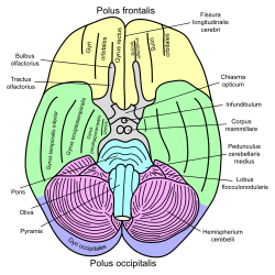
 Basal view of a human brain (Rhinal sulcus not labeled, but is visible posterior to
parahippocampal gyrus.) | |
| Details | |
| Identifiers | |
| Latin | sulcus rhinalis; fissura rhinalis; sulcus rhinicus; fissura rhinica |
| NeuroNames | 41 |
| NeuroLex ID | birnlex_1368 |
| TA98 | A14.1.09.240 |
| TA2 | 5443 |
| FMA | 83746 |
| Anatomical terms of neuroanatomy | |
In the human brain, the entorhinal cortex appears as a longitudinal elevation anterior to the parahippocampal gyrus, with a corresponding internal furrow, the external rhinal sulcus (or rhinal fissure), separating it from the inferiolateral surface of the hemisphere close to the lamina terminalis. It is analogous to the collateral fissure found further caudally in the inferior part of the temporal lobe.
References
![]() This article incorporates text in the
public domain from
page 744 of the 20th edition of
Gray's Anatomy (1918)
This article incorporates text in the
public domain from
page 744 of the 20th edition of
Gray's Anatomy (1918)
External links
| Rhinal sulcus | |
|---|---|
 Basal view of a human brain (Rhinal sulcus not labeled, but is visible posterior to
parahippocampal gyrus.) | |
| Details | |
| Identifiers | |
| Latin | sulcus rhinalis; fissura rhinalis; sulcus rhinicus; fissura rhinica |
| NeuroNames | 41 |
| NeuroLex ID | birnlex_1368 |
| TA98 | A14.1.09.240 |
| TA2 | 5443 |
| FMA | 83746 |
| Anatomical terms of neuroanatomy | |
In the human brain, the entorhinal cortex appears as a longitudinal elevation anterior to the parahippocampal gyrus, with a corresponding internal furrow, the external rhinal sulcus (or rhinal fissure), separating it from the inferiolateral surface of the hemisphere close to the lamina terminalis. It is analogous to the collateral fissure found further caudally in the inferior part of the temporal lobe.
References
![]() This article incorporates text in the
public domain from
page 744 of the 20th edition of
Gray's Anatomy (1918)
This article incorporates text in the
public domain from
page 744 of the 20th edition of
Gray's Anatomy (1918)
External links