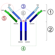
1. Antigen-binding fragment (Fab)
2. Antibody crystallizable region (Fc)
3. Heavy chains
4. Light chains
5. Variable region of the antibody. The paratope is the key-shaped section that makes direct contact with the antigen. [1]
6. Hinge regions
In immunology, a paratope, also known as an antigen-binding site, is the part of an antibody which recognizes and binds to an antigen. [1] [2] It is a small region at the tip of the antibody's antigen-binding fragment and contains parts of the antibody's heavy and light chains. [1] [2] Each paratope is made up of six complementarity-determining regions - three from each of the light and heavy chains - that extend from a fold of anti-parallel beta sheets. [2] Each arm of the Y-shaped antibody has an identical paratope at the end. [2]
Paratopes make up the parts of the B-cell receptor that bind to and make contact with the epitope of an antigen. [2] All the B-cell receptors on any one individual B cell have identical paratopes. [2] The uniqueness of a paratope allows it to bind to only one epitope with high affinity and as a result, each B cell can only respond to one epitope. The paratopes on B-cell receptors binding to their specific epitope is a critical step in the adaptive immune response.
Design of paratopes between species
The design and structure of paratopes can differ greatly between different species. In jawed-vertebrates, V(D)J recombination can result in billions of different paratopes. [3] [4] The number of paratopes, however, is limited by the composition of the V, D, and J genes and the structure of the antibody. [3] Thus, many different species have developed ways to bypass this restriction and increase the diversity of possible paratopes.
In cows, an extra-long complementarity-determining region is considered to have an essential role in diversifying paratopes. [3] [5] Additionally, both chickens and rabbits use gene conversion to increase the number of paratopes that are possible. [3]
References
- ^ a b c Lefranc MP (2013). "Paratope". In Dubitzky W, Wolkenhauer O, Cho KH, Yokota H (eds.). Encyclopedia of Systems Biology. New York, NY: Springer. pp. 1632–1633. doi: 10.1007/978-1-4419-9863-7_673. ISBN 978-1-4419-9863-7.
- ^
a
b
c
d
e
f Punt J, Stranford SA, Jones PP, Owen JA (2019). Kuby immunology (Eighth ed.). New York.
ISBN
978-1-4641-8978-4.
OCLC
1002672752.
{{
cite book}}
: CS1 maint: location missing publisher ( link) - ^ a b c d de los Rios M, Criscitiello MF, Smider VV (August 2015). "Structural and genetic diversity in antibody repertoires from diverse species". Current Opinion in Structural Biology. 33: 27–41. doi: 10.1016/j.sbi.2015.06.002. PMC 7039331. PMID 26188469.
- ^ Litman GW, Rast JP, Fugmann SD (August 2010). "The origins of vertebrate adaptive immunity". Nature Reviews. Immunology. 10 (8): 543–53. doi: 10.1038/nri2807. PMC 2919748. PMID 20651744.
- ^ Wang F, Ekiert DC, Ahmad I, Yu W, Zhang Y, Bazirgan O, et al. (June 2013). "Reshaping antibody diversity". Cell. 153 (6): 1379–93. doi: 10.1016/j.cell.2013.04.049. PMC 4007204. PMID 23746848.