| Middle cerebellar peduncle | |
|---|---|
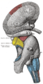
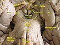
 Dissection showing the projection fibers of the cerebellum. (Middle peduncle labeled at upper right.) | |
| Details | |
| Identifiers | |
| Latin | pedunculus cerebellaris medius |
| MeSH | D065837 |
| NeuroNames | 620 |
| NeuroLex ID | birnlex_1529 |
| TA98 |
A14.1.05.003 A14.1.07.416 |
| TA2 | 5848 |
| FMA | 72515 |
| Anatomical terms of neuroanatomy | |
The middle cerebellar peduncle (or brachium pontis [1]) is a paired structure of the brain. It connects the pons to the cerebellum, with fibres originating from the pontine nucleus and travelling to the opposite hemisphere of the cerebellar cortex. It is supplied by the anterior inferior cerebellar artery (AICA) and branches from the basilar artery. It conveys information from the cerebrum and the pons to the cerebellum.
Structure
The middle cerebellar peduncle is the largest of the three cerebellar peduncles. It connects the pons and cerebellum. It consists almost entirely of fibers passing from the pons to the cerebellum (fibrocerebellar fibers); the fibers arise from the pontine nuclei and decussate within the pons before entering the peduncle [1] to end in the contralateral hemisphere of the cerebellar cortex. [2]
The fibers of the middle cerebellar peduncle are arranged in three fasciculi: superior, inferior, and deep.
- The superior fasciculus, the most superficial, is derived from the upper transverse fibers of the pons; it is directed backward and lateralward superficial to the other two fasciculi, and is distributed mainly to the lobules on the inferior surface of the cerebellar hemisphere and to the parts of the superior surface adjoining the posterior and lateral margins.
- The inferior fasciculus is formed by the lowest transverse fibers of the pons; it passes under cover of the superior fasciculus and is continued downward and backward more or less parallel with it, to be distributed to the folia on the under surface close to the vermis.
- The deep fasciculus comprises most of the deep transverse fibers of the pons. It is at first covered by the superior and inferior fasciculi, but crosses obliquely and appears on the medial side of the superior, from which it receives a bundle; its fibers spread out and pass to the upper anterior cerebellar folia. The fibers of this fasciculus cover those of the inferior cerebellar peduncle.
The trigeminal nerve (CN V) arises from the lateral pons very close to the middle cerebellar peduncle. [3]
Blood supply
The middle cerebellar peduncle is supplied by the anterior inferior cerebellar artery (AICA), as well as smaller branches from the basilar artery. [4]
Function
The middle cerebellar peduncle conveys information from the cerebrum and the pons to the cerebellum. [5]
Clinical significance
Infarction of the anterior inferior cerebellar artery (AICA) can damage the middle cerebellar peduncle. [4] Diffuse intrinsic pontine glioma may spread from the pons into the middle cerebellar peduncle. [6]
Additional images
-
 Scheme showing the connections of the several parts of the brain.
 -
 Superficial dissection of brain-stem. Lateral view.
 -
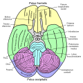
 Hind- and mid-brains; postero-lateral view.
 -
 Upper part of medulla spinalis and hind- and mid-brains; posterior aspect, exposed in situ.
 -
 Basal view of a human brain
 -
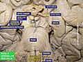
 Dissection of human midbrain with middle cerebellar peduncle labeled.
 -
 Cross section through lower pons showing part of the middle cerebellar peduncle (#19) forming from the convergence of pontocerebellar fibers.
 -
 Middle cerebellar peduncle
 -
 Cerebrum. Deep dissection. Inferior dissection.
 -
 Fourth ventricle. Posterior view.Deep dissection.
 -
 Cerebrum.Inferior view.Deep dissection.
 -
 Cerebrum.Inferior view.Deep dissection.
 -
 Cerebrum.Inferior view.Deep dissection.
 -
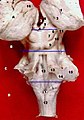
 Cerebellum. Inferior surface.
 -
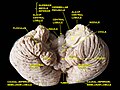 Cerebellum. Inferior surface.
 -
 Cerebellum. Inferior surface.


See also
References
![]() This article incorporates text in the
public domain from the 20th edition of
Gray's Anatomy (1918)
This article incorporates text in the
public domain from the 20th edition of
Gray's Anatomy (1918)
- ^ a b Patestas, Maria A.; Gartner, Leslie P. (2016). A Textbook of Neuroanatomy (2nd ed.). Hoboken, New Jersey: Wiley-Blackwell. p. 290. ISBN 978-1-118-67746-9.
- ^ Voogd, Jan; Ruigrok, Tom J. H. (2012). "15 - Cerebellum and Precerebellar Nuclei". The Human Nervous System (3rd ed.). Academic Press. pp. 471–545. doi: 10.1016/B978-0-12-374236-0.10015-X. ISBN 978-0-12-374236-0.
- ^ Franklin, S. (2017). "5 - The Peripheral and Central Nervous System". Conn's Translational Neuroscience. Academic Press. pp. 113–129. doi: 10.1016/B978-0-12-802381-5.00007-5. ISBN 978-0-12-802381-5.
- ^ a b DeMyer, William (2009). Stroke in Children and Young Adults (2nd ed.). Saunders. pp. 15–68. doi: 10.1016/B978-0-7506-7418-8.00002-1. ISBN 978-0-7506-7418-8.
- ^ Habas, Christophe; Manto, Mario (2018). "14 - Probing the neuroanatomy of the cerebellum using tractography". Handbook of Clinical Neurology. Vol. 154. Elsevier. pp. 235–249. doi: 10.1016/B978-0-444-63956-1.00014-X. ISBN 978-0-444-63956-1. ISSN 0072-9752. PMID 29903442.
- ^ Vitanza, Nicholas A.; Fisher, Paul G.; Deisseroth, Michelle Monje (2017). "128 - Diffuse Intrinsic Pontine Glioma". Swaiman's Pediatric Neurology (6th ed.). Elsevier. pp. 991–994. doi: 10.1016/B978-0-323-37101-8.00128-4. ISBN 978-0-323-37101-8.
External links
- Atlas image: n2a7p4 at the University of Michigan Health System
| Middle cerebellar peduncle | |
|---|---|
|
Dissection showing the projection fibers of the cerebellum. (Middle peduncle labeled at upper right.) | |
| Details | |
| Identifiers | |
| Latin | pedunculus cerebellaris medius |
| MeSH | D065837 |
| NeuroNames | 620 |
| NeuroLex ID | birnlex_1529 |
| TA98 |
A14.1.05.003 A14.1.07.416 |
| TA2 | 5848 |
| FMA | 72515 |
| Anatomical terms of neuroanatomy | |
The middle cerebellar peduncle (or brachium pontis [1]) is a paired structure of the brain. It connects the pons to the cerebellum, with fibres originating from the pontine nucleus and travelling to the opposite hemisphere of the cerebellar cortex. It is supplied by the anterior inferior cerebellar artery (AICA) and branches from the basilar artery. It conveys information from the cerebrum and the pons to the cerebellum.
Structure
The middle cerebellar peduncle is the largest of the three cerebellar peduncles. It connects the pons and cerebellum. It consists almost entirely of fibers passing from the pons to the cerebellum (fibrocerebellar fibers); the fibers arise from the pontine nuclei and decussate within the pons before entering the peduncle [1] to end in the contralateral hemisphere of the cerebellar cortex. [2]
The fibers of the middle cerebellar peduncle are arranged in three fasciculi: superior, inferior, and deep.
- The superior fasciculus, the most superficial, is derived from the upper transverse fibers of the pons; it is directed backward and lateralward superficial to the other two fasciculi, and is distributed mainly to the lobules on the inferior surface of the cerebellar hemisphere and to the parts of the superior surface adjoining the posterior and lateral margins.
- The inferior fasciculus is formed by the lowest transverse fibers of the pons; it passes under cover of the superior fasciculus and is continued downward and backward more or less parallel with it, to be distributed to the folia on the under surface close to the vermis.
- The deep fasciculus comprises most of the deep transverse fibers of the pons. It is at first covered by the superior and inferior fasciculi, but crosses obliquely and appears on the medial side of the superior, from which it receives a bundle; its fibers spread out and pass to the upper anterior cerebellar folia. The fibers of this fasciculus cover those of the inferior cerebellar peduncle.
The trigeminal nerve (CN V) arises from the lateral pons very close to the middle cerebellar peduncle. [3]
Blood supply
The middle cerebellar peduncle is supplied by the anterior inferior cerebellar artery (AICA), as well as smaller branches from the basilar artery. [4]
Function
The middle cerebellar peduncle conveys information from the cerebrum and the pons to the cerebellum. [5]
Clinical significance
Infarction of the anterior inferior cerebellar artery (AICA) can damage the middle cerebellar peduncle. [4] Diffuse intrinsic pontine glioma may spread from the pons into the middle cerebellar peduncle. [6]
Additional images
-
Scheme showing the connections of the several parts of the brain.
-
Superficial dissection of brain-stem. Lateral view.
-
Hind- and mid-brains; postero-lateral view.
-
Upper part of medulla spinalis and hind- and mid-brains; posterior aspect, exposed in situ.
-
Basal view of a human brain
-
Dissection of human midbrain with middle cerebellar peduncle labeled.
-
Cross section through lower pons showing part of the middle cerebellar peduncle (#19) forming from the convergence of pontocerebellar fibers.
-
Middle cerebellar peduncle
-
Cerebrum. Deep dissection. Inferior dissection.
-
Fourth ventricle. Posterior view.Deep dissection.
-
Cerebrum.Inferior view.Deep dissection.
-
Cerebrum.Inferior view.Deep dissection.
-
Cerebrum.Inferior view.Deep dissection.
-
Cerebellum. Inferior surface.
-
Cerebellum. Inferior surface.
-
Cerebellum. Inferior surface.
See also
References
![]() This article incorporates text in the
public domain from the 20th edition of
Gray's Anatomy (1918)
This article incorporates text in the
public domain from the 20th edition of
Gray's Anatomy (1918)
- ^ a b Patestas, Maria A.; Gartner, Leslie P. (2016). A Textbook of Neuroanatomy (2nd ed.). Hoboken, New Jersey: Wiley-Blackwell. p. 290. ISBN 978-1-118-67746-9.
- ^ Voogd, Jan; Ruigrok, Tom J. H. (2012). "15 - Cerebellum and Precerebellar Nuclei". The Human Nervous System (3rd ed.). Academic Press. pp. 471–545. doi: 10.1016/B978-0-12-374236-0.10015-X. ISBN 978-0-12-374236-0.
- ^ Franklin, S. (2017). "5 - The Peripheral and Central Nervous System". Conn's Translational Neuroscience. Academic Press. pp. 113–129. doi: 10.1016/B978-0-12-802381-5.00007-5. ISBN 978-0-12-802381-5.
- ^ a b DeMyer, William (2009). Stroke in Children and Young Adults (2nd ed.). Saunders. pp. 15–68. doi: 10.1016/B978-0-7506-7418-8.00002-1. ISBN 978-0-7506-7418-8.
- ^ Habas, Christophe; Manto, Mario (2018). "14 - Probing the neuroanatomy of the cerebellum using tractography". Handbook of Clinical Neurology. Vol. 154. Elsevier. pp. 235–249. doi: 10.1016/B978-0-444-63956-1.00014-X. ISBN 978-0-444-63956-1. ISSN 0072-9752. PMID 29903442.
- ^ Vitanza, Nicholas A.; Fisher, Paul G.; Deisseroth, Michelle Monje (2017). "128 - Diffuse Intrinsic Pontine Glioma". Swaiman's Pediatric Neurology (6th ed.). Elsevier. pp. 991–994. doi: 10.1016/B978-0-323-37101-8.00128-4. ISBN 978-0-323-37101-8.
External links
- Atlas image: n2a7p4 at the University of Michigan Health System