
Size of this preview:
404 × 599 pixels. Other resolutions:
162 × 240 pixels |
323 × 480 pixels |
801 × 1,188 pixels.
Original file (801 × 1,188 pixels, file size: 176 KB, MIME type: image/png)
| This is a file from the
Wikimedia Commons. Information from its
description page there is shown below. Commons is a freely licensed media file repository. You can help. |
Summary
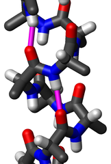
| Description | Close-up sideview of a "stick" model of an alpha helix of poly-alanine using the dihedral angles φ=-60° and ψ=-45° and the Engh&Huber bond geometry. Two hydrogen bonds are highlighted in magenta; the O-H distance is 2.08 Å (208 pm). The PDB file was made by me on 18 October 2006 using my own software and visualized by me using MOLMOL. I release this image under the GFDL. |
| Date | 18 October 2006 (original upload date) |
| Source | No machine-readable source provided. Own work assumed (based on copyright claims). |
| Author | No machine-readable author provided. WillowW assumed (based on copyright claims). |
Licensing
I, the copyright holder of this work, hereby publish it under the following license:

|
Permission is granted to copy, distribute and/or modify this document under the terms of the GNU Free Documentation License, Version 1.2 or any later version published by the Free Software Foundation; with no Invariant Sections, no Front-Cover Texts, and no Back-Cover Texts. A copy of the license is included in the section entitled GNU Free Documentation License. |
| This file is licensed under the Creative Commons Attribution-Share Alike 3.0 Unported license. | ||
| ||
| This licensing tag was added to this file as part of the GFDL licensing update. |
File history
Click on a date/time to view the file as it appeared at that time.
| Date/Time | Thumbnail | Dimensions | User | Comment | |
|---|---|---|---|---|---|
| current | 15:21, 18 October 2006 |
| 801 × 1,188 (176 KB) | WillowW | Same as before, but now showing two hydrogen bonds in magenta, both to the same peptide group. |
| 14:31, 18 October 2006 |
 | 852 × 1,202 (192 KB) | WillowW | Close-up sideview of a "stick" model of an alpha helix of poly-alanine using the dihedral angles φ=-60° and ψ=-45° and the Engh&Huber bond geometry. One hydrogen bond is highlighted in magenta. The PDB file was made by me on 18 October 2006 using my |
File usage
The following pages on the English Wikipedia use this file (pages on other projects are not listed):
Global file usage
The following other wikis use this file:
- Usage on ar.wikipedia.org
- Usage on bs.wikipedia.org
- Usage on ca.wikipedia.org
- Usage on de.wiktionary.org
- Usage on en.wikibooks.org
- Usage on et.wikipedia.org
- Usage on fi.wikipedia.org
- Usage on gl.wikipedia.org
- Usage on he.wikipedia.org
- Usage on hr.wikipedia.org
- Usage on it.wikipedia.org
- Usage on it.wikibooks.org
- Usage on ja.wikipedia.org
- Usage on mk.wikipedia.org
- Usage on pl.wikipedia.org
- Usage on pt.wikipedia.org
- Usage on ru.wikipedia.org
- Вторичная структура
- Альфа-спираль
- Бета-лист
- Карта Рамачандрана
- Мотив «спираль-поворот-спираль»
- Бета-спираль
- Шаблон:Вторичная структура белка
- Бета-шпилька
- Коллагеновая спираль
- Бета-выпуклость
- 310-спираль
- Пи-спираль
- Полипролиновая спираль
- Желейный рулет
- Спиральная катушка
- Сигнал инкапсидации ядра гаммаретровируса
- Супрамолекулярная сборка
- Usage on sh.wikipedia.org
- Usage on simple.wikipedia.org
- Usage on sr.wikipedia.org
- Usage on tr.wikipedia.org
- Usage on uk.wikipedia.org
View more global usage of this file.
Size of this preview:
404 × 599 pixels. Other resolutions:
162 × 240 pixels |
323 × 480 pixels |
801 × 1,188 pixels.
Original file (801 × 1,188 pixels, file size: 176 KB, MIME type: image/png)
| This is a file from the
Wikimedia Commons. Information from its
description page there is shown below. Commons is a freely licensed media file repository. You can help. |
Summary
| Description | Close-up sideview of a "stick" model of an alpha helix of poly-alanine using the dihedral angles φ=-60° and ψ=-45° and the Engh&Huber bond geometry. Two hydrogen bonds are highlighted in magenta; the O-H distance is 2.08 Å (208 pm). The PDB file was made by me on 18 October 2006 using my own software and visualized by me using MOLMOL. I release this image under the GFDL. |
| Date | 18 October 2006 (original upload date) |
| Source | No machine-readable source provided. Own work assumed (based on copyright claims). |
| Author | No machine-readable author provided. WillowW assumed (based on copyright claims). |
Licensing
I, the copyright holder of this work, hereby publish it under the following license:
|
|
Permission is granted to copy, distribute and/or modify this document under the terms of the GNU Free Documentation License, Version 1.2 or any later version published by the Free Software Foundation; with no Invariant Sections, no Front-Cover Texts, and no Back-Cover Texts. A copy of the license is included in the section entitled GNU Free Documentation License. |
| This file is licensed under the Creative Commons Attribution-Share Alike 3.0 Unported license. | ||
| ||
| This licensing tag was added to this file as part of the GFDL licensing update. |
File history
Click on a date/time to view the file as it appeared at that time.
| Date/Time | Thumbnail | Dimensions | User | Comment | |
|---|---|---|---|---|---|
| current | 15:21, 18 October 2006 |
| 801 × 1,188 (176 KB) | WillowW | Same as before, but now showing two hydrogen bonds in magenta, both to the same peptide group. |
| 14:31, 18 October 2006 |
| 852 × 1,202 (192 KB) | WillowW | Close-up sideview of a "stick" model of an alpha helix of poly-alanine using the dihedral angles φ=-60° and ψ=-45° and the Engh&Huber bond geometry. One hydrogen bond is highlighted in magenta. The PDB file was made by me on 18 October 2006 using my |
File usage
The following pages on the English Wikipedia use this file (pages on other projects are not listed):
Global file usage
The following other wikis use this file:
- Usage on ar.wikipedia.org
- Usage on bs.wikipedia.org
- Usage on ca.wikipedia.org
- Usage on de.wiktionary.org
- Usage on en.wikibooks.org
- Usage on et.wikipedia.org
- Usage on fi.wikipedia.org
- Usage on gl.wikipedia.org
- Usage on he.wikipedia.org
- Usage on hr.wikipedia.org
- Usage on it.wikipedia.org
- Usage on it.wikibooks.org
- Usage on ja.wikipedia.org
- Usage on mk.wikipedia.org
- Usage on pl.wikipedia.org
- Usage on pt.wikipedia.org
- Usage on ru.wikipedia.org
- Вторичная структура
- Альфа-спираль
- Бета-лист
- Карта Рамачандрана
- Мотив «спираль-поворот-спираль»
- Бета-спираль
- Шаблон:Вторичная структура белка
- Бета-шпилька
- Коллагеновая спираль
- Бета-выпуклость
- 310-спираль
- Пи-спираль
- Полипролиновая спираль
- Желейный рулет
- Спиральная катушка
- Сигнал инкапсидации ядра гаммаретровируса
- Супрамолекулярная сборка
- Usage on sh.wikipedia.org
- Usage on simple.wikipedia.org
- Usage on sr.wikipedia.org
- Usage on tr.wikipedia.org
- Usage on uk.wikipedia.org
View more global usage of this file.