Size of this preview:
800 × 358 pixels. Other resolutions:
320 × 143 pixels |
640 × 286 pixels |
1,024 × 458 pixels |
1,280 × 572 pixels |
2,560 × 1,145 pixels |
6,274 × 2,806 pixels.
Original file (6,274 × 2,806 pixels, file size: 757 KB, MIME type: image/png)
| This is a file from the
Wikimedia Commons. Information from its
description page there is shown below. Commons is a freely licensed media file repository. You can help. |
Summary
| Description |
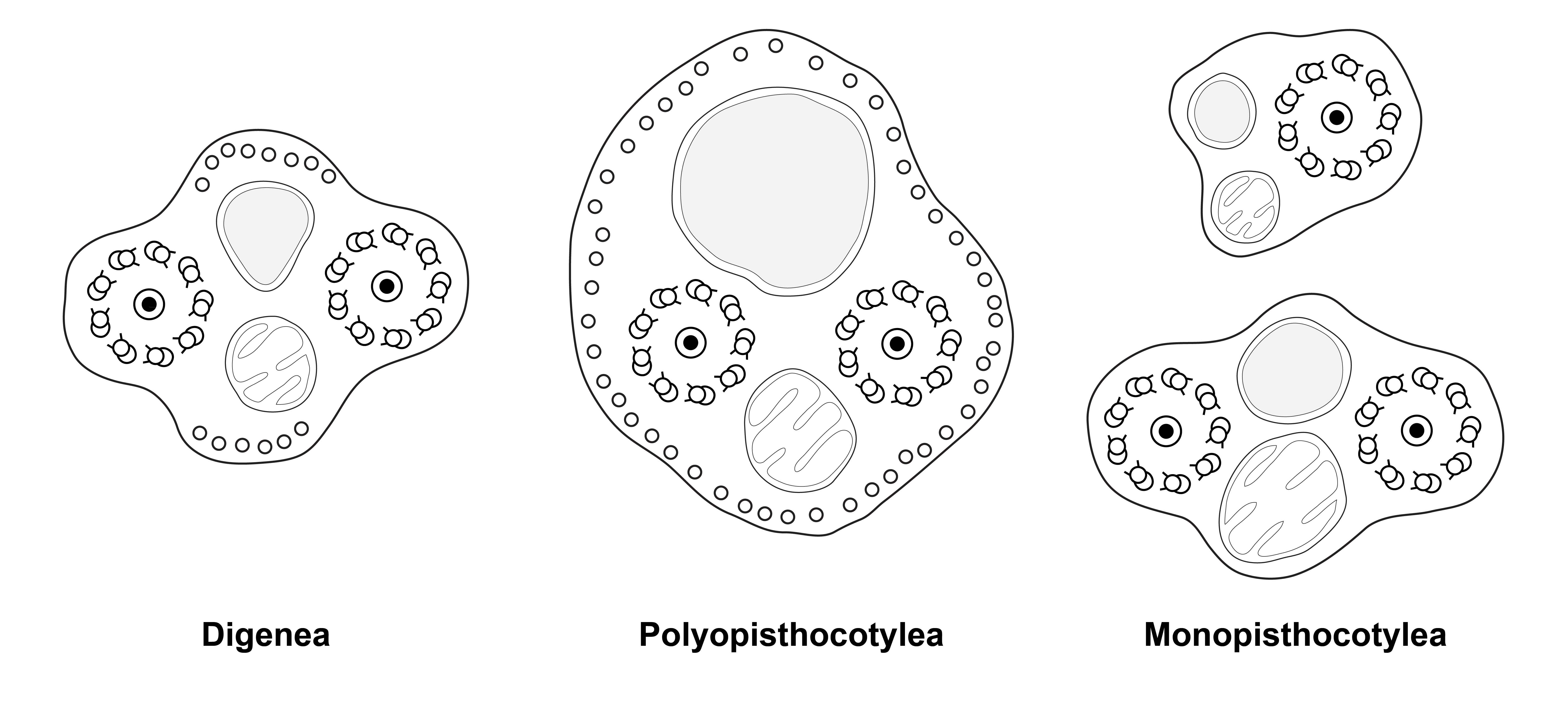
English: Figure 16 of the paper. Diagrams of spermatozoa (redrawn from Justine, 1991). Caption adapted from original caption of figure. Diagrams were drawn from original micrographs of transverse sections. Digenea. Dorsal and ventral microtubules are present (proposed as a synapomorphy for the Cercomeridea). There are no lateral microtubules (symplesiomorphic compared with the synapomorphy for the polyopisthocotylean Monogenea). Polyopisthocotylea. Dorsal and ventral microtubules are present (synapomorphy for Cercomeridea). Note the presence of lateral microtubules (proposed as a synapomorphy for the Polyopisthocotylea). Monopisthocotylea (uniflagellate and biflagellate). Microtubules are absent from the principal region of the spermatozoon, which is interpreted as (i) the absence of dorsal and ventral microtubules, a reversal of the synapomorphy for the Cercomeridea; and (ii) the absence of lateral microtubules, the symplesiomorphic state versus the synapomorphy for the Polyopisthocotylea. |
| Date | |
| Source | (2018). " Spermiogenesis and spermatozoon ultrastructure in basal polyopisthocotylean monogeneans, Hexabothriidae and Chimaericolidae, and their significance for the phylogeny of the Monogenea". Parasite 25: 7. DOI: 10.1051/parasite/2018007. ISSN 1776-1042. |
| Author | Jean-Lou Justine and Larisa G. Poddubnaya |
Licensing
This file is licensed under the
Creative Commons
Attribution 4.0 International license.
|
 This file was published in the scientific journal
Parasite.
Their website states that all content of the journal including and after 2013 is published under the Creative Commons Attribution 4.0 license.
|
File history
Click on a date/time to view the file as it appeared at that time.
| Date/Time | Thumbnail | Dimensions | User | Comment | |
|---|---|---|---|---|---|
| current | 16:58, 15 February 2018 |
| 6,274 × 2,806 (757 KB) | Jeanloujustine | User created page with UploadWizard |
File usage
The following pages on the English Wikipedia use this file (pages on other projects are not listed):
Global file usage
The following other wikis use this file:
- Usage on ca.wikipedia.org
- Usage on fa.wikipedia.org
- Usage on tr.wikipedia.org
- Usage on www.wikidata.org
Metadata
Size of this preview:
800 × 358 pixels. Other resolutions:
320 × 143 pixels |
640 × 286 pixels |
1,024 × 458 pixels |
1,280 × 572 pixels |
2,560 × 1,145 pixels |
6,274 × 2,806 pixels.
Original file (6,274 × 2,806 pixels, file size: 757 KB, MIME type: image/png)
| This is a file from the
Wikimedia Commons. Information from its
description page there is shown below. Commons is a freely licensed media file repository. You can help. |
Summary
| Description |
English: Figure 16 of the paper. Diagrams of spermatozoa (redrawn from Justine, 1991). Caption adapted from original caption of figure. Diagrams were drawn from original micrographs of transverse sections. Digenea. Dorsal and ventral microtubules are present (proposed as a synapomorphy for the Cercomeridea). There are no lateral microtubules (symplesiomorphic compared with the synapomorphy for the polyopisthocotylean Monogenea). Polyopisthocotylea. Dorsal and ventral microtubules are present (synapomorphy for Cercomeridea). Note the presence of lateral microtubules (proposed as a synapomorphy for the Polyopisthocotylea). Monopisthocotylea (uniflagellate and biflagellate). Microtubules are absent from the principal region of the spermatozoon, which is interpreted as (i) the absence of dorsal and ventral microtubules, a reversal of the synapomorphy for the Cercomeridea; and (ii) the absence of lateral microtubules, the symplesiomorphic state versus the synapomorphy for the Polyopisthocotylea. |
| Date | |
| Source | (2018). " Spermiogenesis and spermatozoon ultrastructure in basal polyopisthocotylean monogeneans, Hexabothriidae and Chimaericolidae, and their significance for the phylogeny of the Monogenea". Parasite 25: 7. DOI: 10.1051/parasite/2018007. ISSN 1776-1042. |
| Author | Jean-Lou Justine and Larisa G. Poddubnaya |
Licensing
This file is licensed under the
Creative Commons
Attribution 4.0 International license.
|
This file was published in the scientific journal
Parasite.
Their website states that all content of the journal including and after 2013 is published under the Creative Commons Attribution 4.0 license.
|
File history
Click on a date/time to view the file as it appeared at that time.
| Date/Time | Thumbnail | Dimensions | User | Comment | |
|---|---|---|---|---|---|
| current | 16:58, 15 February 2018 |
| 6,274 × 2,806 (757 KB) | Jeanloujustine | User created page with UploadWizard |
File usage
The following pages on the English Wikipedia use this file (pages on other projects are not listed):
Global file usage
The following other wikis use this file:
- Usage on ca.wikipedia.org
- Usage on fa.wikipedia.org
- Usage on tr.wikipedia.org
- Usage on www.wikidata.org