
Size of this preview:
593 × 599 pixels. Other resolutions:
237 × 240 pixels |
475 × 480 pixels |
760 × 768 pixels |
1,013 × 1,024 pixels |
1,768 × 1,787 pixels.
Original file (1,768 × 1,787 pixels, file size: 4.03 MB, MIME type: image/jpeg)
| This is a file from the
Wikimedia Commons. Information from its
description page there is shown below. Commons is a freely licensed media file repository. You can help. |
Summary
| Description |
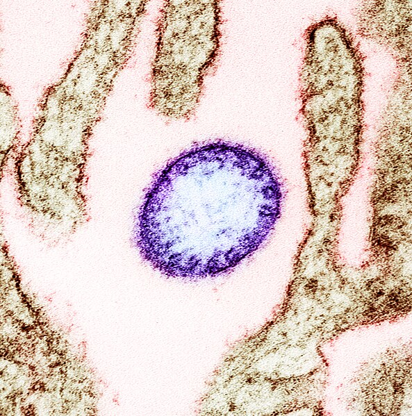
English: Colorized transmission electron micrograph of a mature extracellular Nipah Virus particle (purple) near the periphery of an infected VERO cell (brown). Image captured and color-enhanced at the NIAID Integrated Research Facility in Fort Detrick, Maryland. Credit: NIAID |
| Date | |
| Source | https://www.flickr.com/photos/niaid/43112803134 |
| Author | NIAID |
Licensing
| This image was originally posted to Flickr by NIAID at https://flickr.com/photos/54591706@N02/43112803134 ( archive). It was reviewed on 18 June 2019 by FlickreviewR 2 and was confirmed to be licensed under the terms of the cc-by-2.0. |
This file is licensed under the
Creative Commons
Attribution 2.0 Generic license.
- You are free:
- to share – to copy, distribute and transmit the work
- to remix – to adapt the work
- Under the following conditions:
- attribution – You must give appropriate credit, provide a link to the license, and indicate if changes were made. You may do so in any reasonable manner, but not in any way that suggests the licensor endorses you or your use.
File history
Click on a date/time to view the file as it appeared at that time.
| Date/Time | Thumbnail | Dimensions | User | Comment | |
|---|---|---|---|---|---|
| current | 21:20, 18 June 2019 |
| 1,768 × 1,787 (4.03 MB) | Bob Saint Clar | User created page with UploadWizard |
File usage
The following pages on the English Wikipedia use this file (pages on other projects are not listed):
Global file usage
The following other wikis use this file:
- Usage on arz.wikipedia.org
- Usage on bcl.wikipedia.org
- Usage on bn.wikipedia.org
- Usage on ca.wikipedia.org
- Usage on da.wikipedia.org
- Usage on de.wikipedia.org
- Usage on en.wiktionary.org
- Usage on fa.wikipedia.org
- Usage on fr.wikipedia.org
- Usage on ha.wikipedia.org
- Usage on ia.wikipedia.org
- Usage on it.wikipedia.org
- Usage on ja.wikipedia.org
- Usage on ml.wikipedia.org
- Usage on mr.wikipedia.org
- Usage on pt.wikipedia.org
- Usage on sv.wikipedia.org
- Usage on uk.wikipedia.org
- Usage on www.wikidata.org
Metadata
Size of this preview:
593 × 599 pixels. Other resolutions:
237 × 240 pixels |
475 × 480 pixels |
760 × 768 pixels |
1,013 × 1,024 pixels |
1,768 × 1,787 pixels.
Original file (1,768 × 1,787 pixels, file size: 4.03 MB, MIME type: image/jpeg)
| This is a file from the
Wikimedia Commons. Information from its
description page there is shown below. Commons is a freely licensed media file repository. You can help. |
Summary
| Description |
English: Colorized transmission electron micrograph of a mature extracellular Nipah Virus particle (purple) near the periphery of an infected VERO cell (brown). Image captured and color-enhanced at the NIAID Integrated Research Facility in Fort Detrick, Maryland. Credit: NIAID |
| Date | |
| Source | https://www.flickr.com/photos/niaid/43112803134 |
| Author | NIAID |
Licensing
| This image was originally posted to Flickr by NIAID at https://flickr.com/photos/54591706@N02/43112803134 ( archive). It was reviewed on 18 June 2019 by FlickreviewR 2 and was confirmed to be licensed under the terms of the cc-by-2.0. |
This file is licensed under the
Creative Commons
Attribution 2.0 Generic license.
- You are free:
- to share – to copy, distribute and transmit the work
- to remix – to adapt the work
- Under the following conditions:
- attribution – You must give appropriate credit, provide a link to the license, and indicate if changes were made. You may do so in any reasonable manner, but not in any way that suggests the licensor endorses you or your use.
File history
Click on a date/time to view the file as it appeared at that time.
| Date/Time | Thumbnail | Dimensions | User | Comment | |
|---|---|---|---|---|---|
| current | 21:20, 18 June 2019 |
| 1,768 × 1,787 (4.03 MB) | Bob Saint Clar | User created page with UploadWizard |
File usage
The following pages on the English Wikipedia use this file (pages on other projects are not listed):
Global file usage
The following other wikis use this file:
- Usage on arz.wikipedia.org
- Usage on bcl.wikipedia.org
- Usage on bn.wikipedia.org
- Usage on ca.wikipedia.org
- Usage on da.wikipedia.org
- Usage on de.wikipedia.org
- Usage on en.wiktionary.org
- Usage on fa.wikipedia.org
- Usage on fr.wikipedia.org
- Usage on ha.wikipedia.org
- Usage on ia.wikipedia.org
- Usage on it.wikipedia.org
- Usage on ja.wikipedia.org
- Usage on ml.wikipedia.org
- Usage on mr.wikipedia.org
- Usage on pt.wikipedia.org
- Usage on sv.wikipedia.org
- Usage on uk.wikipedia.org
- Usage on www.wikidata.org