
Size of this preview:
765 × 599 pixels. Other resolutions:
306 × 240 pixels |
613 × 480 pixels |
980 × 768 pixels |
1,206 × 945 pixels.
Original file (1,206 × 945 pixels, file size: 607 KB, MIME type: image/jpeg)
| This is a file from the
Wikimedia Commons. Information from its
description page there is shown below. Commons is a freely licensed media file repository. You can help. |
Summary
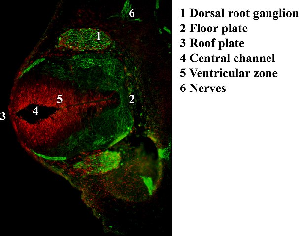
| Description | Antibody stain against Neurofilament (green) and Ki 67 (red) in a Mouse embryo at day 12.5 after fertilization. Shown is the dorsal root ganglion (green ellipsoid regions where cells express neurofilament) and the ventricular zone (red region where cells proliferate) as well as the neural tube with roof and floor plate. |
| Date | 04.05.2007 |
| Source | Own work |
| Author | Hannes Röst |
Licensing
| I, the copyright holder of this work, release this work into the
public domain. This applies worldwide. In some countries this may not be legally possible; if so: I grant anyone the right to use this work for any purpose, without any conditions, unless such conditions are required by law. |
File history
Click on a date/time to view the file as it appeared at that time.
| Date/Time | Thumbnail | Dimensions | User | Comment | |
|---|---|---|---|---|---|
| current | 23:30, 1 November 2007 |
| 1,206 × 945 (607 KB) | Hannes Röst | == Summary == {{Information |Description=Antibody stain against Neurofilament (green) and Ki 67 (red) in a Mouse embryo at day 12.5 after fertilization. Shown is the dorsal root ganglion (green ellipsoid regions where cells express neurofilament) and the |
| 22:03, 16 May 2007 |
 | 1,181 × 945 (149 KB) | Hannes Röst | {{Information |Description=Antibody stain against Neurofilament (green) and Ki 67 (red) in a Mouse embryo. Shown is the neural tube development. |Source=self-made |Date=04.05.2007 |Author= User:Hannes Röst }} |
File usage
The following pages on the English Wikipedia use this file (pages on other projects are not listed):
Global file usage
The following other wikis use this file:
- Usage on ca.wikipedia.org
- Usage on es.wikipedia.org
- Usage on fr.wikipedia.org
- Usage on gl.wikipedia.org
- Usage on ja.wikipedia.org
- Usage on pl.wikipedia.org
Metadata
Size of this preview:
765 × 599 pixels. Other resolutions:
306 × 240 pixels |
613 × 480 pixels |
980 × 768 pixels |
1,206 × 945 pixels.
Original file (1,206 × 945 pixels, file size: 607 KB, MIME type: image/jpeg)
| This is a file from the
Wikimedia Commons. Information from its
description page there is shown below. Commons is a freely licensed media file repository. You can help. |
Summary
| Description | Antibody stain against Neurofilament (green) and Ki 67 (red) in a Mouse embryo at day 12.5 after fertilization. Shown is the dorsal root ganglion (green ellipsoid regions where cells express neurofilament) and the ventricular zone (red region where cells proliferate) as well as the neural tube with roof and floor plate. |
| Date | 04.05.2007 |
| Source | Own work |
| Author | Hannes Röst |
Licensing
| I, the copyright holder of this work, release this work into the
public domain. This applies worldwide. In some countries this may not be legally possible; if so: I grant anyone the right to use this work for any purpose, without any conditions, unless such conditions are required by law. |
File history
Click on a date/time to view the file as it appeared at that time.
| Date/Time | Thumbnail | Dimensions | User | Comment | |
|---|---|---|---|---|---|
| current | 23:30, 1 November 2007 |
| 1,206 × 945 (607 KB) | Hannes Röst | == Summary == {{Information |Description=Antibody stain against Neurofilament (green) and Ki 67 (red) in a Mouse embryo at day 12.5 after fertilization. Shown is the dorsal root ganglion (green ellipsoid regions where cells express neurofilament) and the |
| 22:03, 16 May 2007 |
| 1,181 × 945 (149 KB) | Hannes Röst | {{Information |Description=Antibody stain against Neurofilament (green) and Ki 67 (red) in a Mouse embryo. Shown is the neural tube development. |Source=self-made |Date=04.05.2007 |Author= User:Hannes Röst }} |
File usage
The following pages on the English Wikipedia use this file (pages on other projects are not listed):
Global file usage
The following other wikis use this file:
- Usage on ca.wikipedia.org
- Usage on es.wikipedia.org
- Usage on fr.wikipedia.org
- Usage on gl.wikipedia.org
- Usage on ja.wikipedia.org
- Usage on pl.wikipedia.org