
Original file (1,115 × 491 pixels, file size: 192 KB, MIME type: image/png)
| This is a file from the
Wikimedia Commons. Information from its
description page there is shown below. Commons is a freely licensed media file repository. You can help. |
Summary
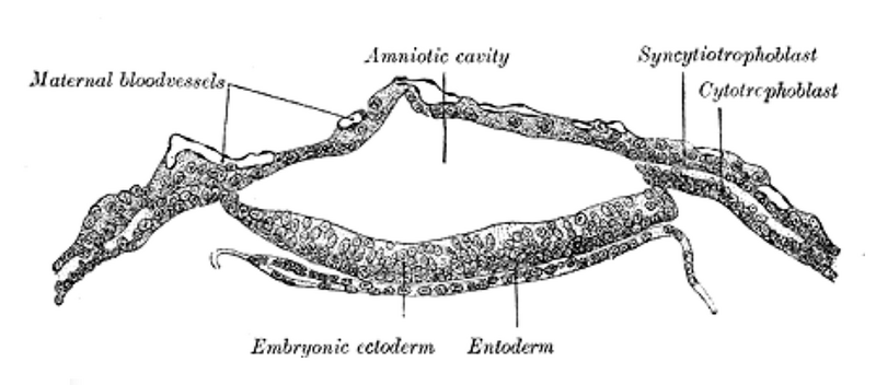
| Description | Section through embryonic area of Vespertilio murinus to show the formation of the amniotic cavity. (After van Beneden.) | ||||||||||||||||||||
| Plate | 12 | ||||||||||||||||||||
| Date | before 1858 | ||||||||||||||||||||
| Source |
|
||||||||||||||||||||
| Author |
|
||||||||||||||||||||
.jpg)
Book
|
Henry Gray:
Gray's Anatomy (20th edition)
|
|||||||||||||||||||||||
|---|---|---|---|---|---|---|---|---|---|---|---|---|---|---|---|---|---|---|---|---|---|---|---|
| Author |
|
-_Title_page.png) | |||||||||||||||||||||
| Editor |
Revised by Warren H. Lewis |
||||||||||||||||||||||
| Illustrator |
|
||||||||||||||||||||||
| Title | |||||||||||||||||||||||
| Edition |
20
|
||||||||||||||||||||||
| Publisher | |||||||||||||||||||||||
| Object type |
version, edition or translation
|
||||||||||||||||||||||
| Page overview | list of all the plates | ||||||||||||||||||||||
| Language |
English
|
||||||||||||||||||||||
| Publication date |
1918
|
||||||||||||||||||||||
| Place of publication |
Philadelphia /
New York City
|
||||||||||||||||||||||
| Source | Bartleby | ||||||||||||||||||||||

Licensing
This image is in the
public domain because it is a mere mechanical scan or photocopy of a public domain original, or – from the available evidence – is so similar to such a scan or photocopy that no copyright protection can be expected to arise. The original itself is in the public domain for the following reason:
This tag is designed for use where there may be a need to assert that any enhancements (eg brightness, contrast, colour-matching, sharpening) are in themselves insufficiently creative to generate a new copyright. It can be used where it is unknown whether any enhancements have been made, as well as when the enhancements are clear but insufficient. For known raw unenhanced scans you can use an appropriate {{PD-old}} tag instead. For usage, see Commons:When to use the PD-scan tag.  | ||||
File history
Click on a date/time to view the file as it appeared at that time.
| Date/Time | Thumbnail | Dimensions | User | Comment | |
|---|---|---|---|---|---|
| current | 20:00, 14 June 2024 |
| 1,115 × 491 (192 KB) | Rasbak | |
| 19:18, 23 January 2007 |
| 450 × 181 (12 KB) | Pngbot | optimized with optipng | |
| 11:43, 18 May 2006 |
| 450 × 181 (12 KB) | File Upload Bot (Magnus Manske) | {{Information| |Description= {{Gray's Anatomy plate|Section through embryonic area of Vespertilio murinus to show the formation of the amniotic cavity. (After van Beneden.)}} |Source=Originally from [http://en.wikipedia.org en.wikipedia]; description pag |
File usage
Global file usage
The following other wikis use this file:
- Usage on ar.wikipedia.org
- Usage on bg.wikipedia.org
- Usage on de.wikibooks.org
- Usage on es.wikipedia.org
- Usage on fa.wikipedia.org
- Usage on gl.wikipedia.org
- Usage on sk.wikipedia.org
- Usage on vi.wikipedia.org
- Usage on zh.wikipedia.org
Metadata
Original file (1,115 × 491 pixels, file size: 192 KB, MIME type: image/png)
| This is a file from the
Wikimedia Commons. Information from its
description page there is shown below. Commons is a freely licensed media file repository. You can help. |
Summary
| Description | Section through embryonic area of Vespertilio murinus to show the formation of the amniotic cavity. (After van Beneden.) | ||||||||||||||||||||
| Plate | 12 | ||||||||||||||||||||
| Date | before 1858 | ||||||||||||||||||||
| Source |
|
||||||||||||||||||||
| Author |
|
||||||||||||||||||||
Book
|
Henry Gray:
Gray's Anatomy (20th edition)
|
|||||||||||||||||||||||
|---|---|---|---|---|---|---|---|---|---|---|---|---|---|---|---|---|---|---|---|---|---|---|---|
| Author |
|
| |||||||||||||||||||||
| Editor |
Revised by Warren H. Lewis |
||||||||||||||||||||||
| Illustrator |
|
||||||||||||||||||||||
| Title | |||||||||||||||||||||||
| Edition |
20
|
||||||||||||||||||||||
| Publisher | |||||||||||||||||||||||
| Object type |
version, edition or translation
|
||||||||||||||||||||||
| Page overview | list of all the plates | ||||||||||||||||||||||
| Language |
English
|
||||||||||||||||||||||
| Publication date |
1918
|
||||||||||||||||||||||
| Place of publication |
Philadelphia /
New York City
|
||||||||||||||||||||||
| Source | Bartleby | ||||||||||||||||||||||
Licensing
This image is in the
public domain because it is a mere mechanical scan or photocopy of a public domain original, or – from the available evidence – is so similar to such a scan or photocopy that no copyright protection can be expected to arise. The original itself is in the public domain for the following reason:
This tag is designed for use where there may be a need to assert that any enhancements (eg brightness, contrast, colour-matching, sharpening) are in themselves insufficiently creative to generate a new copyright. It can be used where it is unknown whether any enhancements have been made, as well as when the enhancements are clear but insufficient. For known raw unenhanced scans you can use an appropriate {{PD-old}} tag instead. For usage, see Commons:When to use the PD-scan tag. | ||||
File history
Click on a date/time to view the file as it appeared at that time.
| Date/Time | Thumbnail | Dimensions | User | Comment | |
|---|---|---|---|---|---|
| current | 20:00, 14 June 2024 |
| 1,115 × 491 (192 KB) | Rasbak | |
| 19:18, 23 January 2007 |
| 450 × 181 (12 KB) | Pngbot | optimized with optipng | |
| 11:43, 18 May 2006 |
| 450 × 181 (12 KB) | File Upload Bot (Magnus Manske) | {{Information| |Description= {{Gray's Anatomy plate|Section through embryonic area of Vespertilio murinus to show the formation of the amniotic cavity. (After van Beneden.)}} |Source=Originally from [http://en.wikipedia.org en.wikipedia]; description pag |
File usage
Global file usage
The following other wikis use this file:
- Usage on ar.wikipedia.org
- Usage on bg.wikipedia.org
- Usage on de.wikibooks.org
- Usage on es.wikipedia.org
- Usage on fa.wikipedia.org
- Usage on gl.wikipedia.org
- Usage on sk.wikipedia.org
- Usage on vi.wikipedia.org
- Usage on zh.wikipedia.org