
Size of this PNG preview of this TIF file:
747 × 599 pixels. Other resolutions:
299 × 240 pixels |
598 × 480 pixels |
957 × 768 pixels |
1,276 × 1,024 pixels |
2,553 × 2,048 pixels |
2,752 × 2,208 pixels.
Original file (2,752 × 2,208 pixels, file size: 17.8 MB, MIME type: image/tiff)
| This is a file from the
Wikimedia Commons. Information from its
description page there is shown below. Commons is a freely licensed media file repository. You can help. |
Summary
| Description |
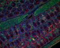
English: Immunofluorescence staining showing proximal part of the large intestine from 8 weeks old mouse. Cells are stained for beta-actin (red), proliferating cell nuclear antigen (green) and nucleus (blue). This image was taken using a Zeiss microscope (20x objective). |
| Date | |
| Source | Own work |
| Author | Abhimanu.pandey |
Licensing
I, the copyright holder of this work, hereby publish it under the following license:
This file is licensed under the
Creative Commons
Attribution 4.0 International license.
- You are free:
- to share – to copy, distribute and transmit the work
- to remix – to adapt the work
- Under the following conditions:
- attribution – You must give appropriate credit, provide a link to the license, and indicate if changes were made. You may do so in any reasonable manner, but not in any way that suggests the licensor endorses you or your use.
| This image was uploaded as part of Wiki Science Competition 2019. |
File history
Click on a date/time to view the file as it appeared at that time.
| Date/Time | Thumbnail | Dimensions | User | Comment | |
|---|---|---|---|---|---|
| current | 00:19, 7 December 2019 |
 | 2,752 × 2,208 (17.8 MB) | Abhimanu.pandey | User created page with UploadWizard |
File usage
The following pages on the English Wikipedia use this file (pages on other projects are not listed):
- User:Ambrosia10/Archive 4
- User talk:Abalg
- User talk:Ambrosia10/Archive 4
- User talk:Awkwafaba
- User talk:Cwmhiraeth/Archive 21
- User talk:Cygnis insignis/Archive 5
- User talk:Deuterostome
- User talk:Dick Bos/archive2020
- User talk:Donald Albury/Archive 13
- User talk:Elmidae/Archive 10
- User talk:Enwebb/Archive 4
- User talk:Epipelagic/Archive 13
- User talk:Evolution and evolvability/Archive 2020
- User talk:Froggydame
- User talk:FunkMonk/Archive 24
- User talk:GreatSculptorIthas
- User talk:Guettarda/Archive22
- User talk:JarrahTree/Archive 61
- User talk:Jens Lallensack/Archive 1
- User talk:JoJan/Archive 25
- User talk:Jowaninpensans
- User talk:Kiwikiu
- User talk:LittleLazyLass
- User talk:Maias
- User talk:MarkZusab/archive6
- User talk:Micromesistius
- User talk:Pvmoutside/Archive 4
- User talk:Quetzal1964/Archive 2
- User talk:Sbbarker19
- User talk:SchreiberBike/Archive 6
- User talk:SilverTiger12
- User talk:Slate Weasel
- User talk:Starsandwhales/2020
- User talk:William Avery/Archive 12
- Wikipedia:WikiProject Tree of Life/Newsletter/011
Metadata
Size of this PNG preview of this TIF file:
747 × 599 pixels. Other resolutions:
299 × 240 pixels |
598 × 480 pixels |
957 × 768 pixels |
1,276 × 1,024 pixels |
2,553 × 2,048 pixels |
2,752 × 2,208 pixels.
Original file (2,752 × 2,208 pixels, file size: 17.8 MB, MIME type: image/tiff)
| This is a file from the
Wikimedia Commons. Information from its
description page there is shown below. Commons is a freely licensed media file repository. You can help. |
Summary
| Description |
English: Immunofluorescence staining showing proximal part of the large intestine from 8 weeks old mouse. Cells are stained for beta-actin (red), proliferating cell nuclear antigen (green) and nucleus (blue). This image was taken using a Zeiss microscope (20x objective). |
| Date | |
| Source | Own work |
| Author | Abhimanu.pandey |
Licensing
I, the copyright holder of this work, hereby publish it under the following license:
This file is licensed under the
Creative Commons
Attribution 4.0 International license.
- You are free:
- to share – to copy, distribute and transmit the work
- to remix – to adapt the work
- Under the following conditions:
- attribution – You must give appropriate credit, provide a link to the license, and indicate if changes were made. You may do so in any reasonable manner, but not in any way that suggests the licensor endorses you or your use.
| This image was uploaded as part of Wiki Science Competition 2019. |
File history
Click on a date/time to view the file as it appeared at that time.
| Date/Time | Thumbnail | Dimensions | User | Comment | |
|---|---|---|---|---|---|
| current | 00:19, 7 December 2019 |
| 2,752 × 2,208 (17.8 MB) | Abhimanu.pandey | User created page with UploadWizard |
File usage
The following pages on the English Wikipedia use this file (pages on other projects are not listed):
- User:Ambrosia10/Archive 4
- User talk:Abalg
- User talk:Ambrosia10/Archive 4
- User talk:Awkwafaba
- User talk:Cwmhiraeth/Archive 21
- User talk:Cygnis insignis/Archive 5
- User talk:Deuterostome
- User talk:Dick Bos/archive2020
- User talk:Donald Albury/Archive 13
- User talk:Elmidae/Archive 10
- User talk:Enwebb/Archive 4
- User talk:Epipelagic/Archive 13
- User talk:Evolution and evolvability/Archive 2020
- User talk:Froggydame
- User talk:FunkMonk/Archive 24
- User talk:GreatSculptorIthas
- User talk:Guettarda/Archive22
- User talk:JarrahTree/Archive 61
- User talk:Jens Lallensack/Archive 1
- User talk:JoJan/Archive 25
- User talk:Jowaninpensans
- User talk:Kiwikiu
- User talk:LittleLazyLass
- User talk:Maias
- User talk:MarkZusab/archive6
- User talk:Micromesistius
- User talk:Pvmoutside/Archive 4
- User talk:Quetzal1964/Archive 2
- User talk:Sbbarker19
- User talk:SchreiberBike/Archive 6
- User talk:SilverTiger12
- User talk:Slate Weasel
- User talk:Starsandwhales/2020
- User talk:William Avery/Archive 12
- Wikipedia:WikiProject Tree of Life/Newsletter/011