
Chain_of_conidia_of_an_Alternaria_sp._fungus_PHIL_3963_lores.jpg (700 × 457 pixels, file size: 22 KB, MIME type: image/jpeg)
| This is a file from the
Wikimedia Commons. Information from its
description page there is shown below. Commons is a freely licensed media file repository. You can help. |
| Description |
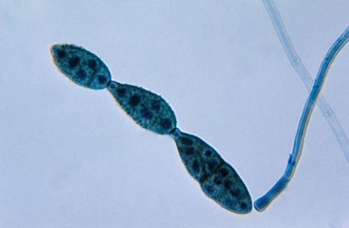
ID#: 3963 Description: This photomicrograph shows a chain of conidia of a Alternaria sp. fungus, which can be a cause of phaeohyphomycosis. The spores of Alternaria sp. fungi are multi-cellular, pigmented, and are produced in straight chains, or branching chains. The end of the conidium nearest the conidiophore is round as it tapers towards its apex, imparting a beak-like appearance. Content Providers(s): CDC/Dr. Lucille K. Georg Creation Date: 1955 Copyright Restrictions: None - This image is in the public domain and thus free of any copyright restrictions. As a matter of courtesy we request that the content provider be credited and notified in any public or private usage of this image. |
|||
| Source | http://phil.cdc.gov/phil_images/20030612/9/PHIL_3963_lores.jpg | |||
| Author | ||||
| Permission ( Reusing this file) |
|
File history
Click on a date/time to view the file as it appeared at that time.
| Date/Time | Thumbnail | Dimensions | User | Comment | |
|---|---|---|---|---|---|
| current | 20:30, 6 May 2006 |
| 700 × 457 (22 KB) | Patho | {{Information| |Description=ID#: 3963 Description: This photomicrograph shows a chain of conidia of a Alternaria sp. fungus, which can be a cause of phaeohyphomycosis. The spores of Alternaria sp. fungi are multi-cellular, pigmented, and are produced in |
File usage
Global file usage
The following other wikis use this file:
- Usage on ar.wikipedia.org
- Usage on arz.wikipedia.org
- Usage on ca.wikipedia.org
- Usage on ceb.wikipedia.org
- Usage on de.wikibooks.org
- Usage on eo.wikipedia.org
- Usage on es.wikipedia.org
- Usage on fa.wikipedia.org
- Usage on fr.wikipedia.org
- Usage on gl.wikipedia.org
- Usage on hy.wikipedia.org
- Usage on ia.wikipedia.org
- Usage on id.wikipedia.org
- Usage on it.wikipedia.org
- Usage on ja.wikipedia.org
- Usage on nl.wikipedia.org
- Usage on om.wikipedia.org
- Usage on pt.wikipedia.org
- Usage on ro.wikipedia.org
- Usage on ro.wiktionary.org
- Usage on ru.wikipedia.org
- Usage on simple.wikipedia.org
- Usage on sv.wikipedia.org
- Usage on tr.wikipedia.org
- Usage on uk.wikipedia.org
View more global usage of this file.
Chain_of_conidia_of_an_Alternaria_sp._fungus_PHIL_3963_lores.jpg (700 × 457 pixels, file size: 22 KB, MIME type: image/jpeg)
| This is a file from the
Wikimedia Commons. Information from its
description page there is shown below. Commons is a freely licensed media file repository. You can help. |
| Description |
ID#: 3963 Description: This photomicrograph shows a chain of conidia of a Alternaria sp. fungus, which can be a cause of phaeohyphomycosis. The spores of Alternaria sp. fungi are multi-cellular, pigmented, and are produced in straight chains, or branching chains. The end of the conidium nearest the conidiophore is round as it tapers towards its apex, imparting a beak-like appearance. Content Providers(s): CDC/Dr. Lucille K. Georg Creation Date: 1955 Copyright Restrictions: None - This image is in the public domain and thus free of any copyright restrictions. As a matter of courtesy we request that the content provider be credited and notified in any public or private usage of this image. |
|||
| Source | http://phil.cdc.gov/phil_images/20030612/9/PHIL_3963_lores.jpg | |||
| Author | ||||
| Permission ( Reusing this file) |
|
File history
Click on a date/time to view the file as it appeared at that time.
| Date/Time | Thumbnail | Dimensions | User | Comment | |
|---|---|---|---|---|---|
| current | 20:30, 6 May 2006 |
| 700 × 457 (22 KB) | Patho | {{Information| |Description=ID#: 3963 Description: This photomicrograph shows a chain of conidia of a Alternaria sp. fungus, which can be a cause of phaeohyphomycosis. The spores of Alternaria sp. fungi are multi-cellular, pigmented, and are produced in |
File usage
Global file usage
The following other wikis use this file:
- Usage on ar.wikipedia.org
- Usage on arz.wikipedia.org
- Usage on ca.wikipedia.org
- Usage on ceb.wikipedia.org
- Usage on de.wikibooks.org
- Usage on eo.wikipedia.org
- Usage on es.wikipedia.org
- Usage on fa.wikipedia.org
- Usage on fr.wikipedia.org
- Usage on gl.wikipedia.org
- Usage on hy.wikipedia.org
- Usage on ia.wikipedia.org
- Usage on id.wikipedia.org
- Usage on it.wikipedia.org
- Usage on ja.wikipedia.org
- Usage on nl.wikipedia.org
- Usage on om.wikipedia.org
- Usage on pt.wikipedia.org
- Usage on ro.wikipedia.org
- Usage on ro.wiktionary.org
- Usage on ru.wikipedia.org
- Usage on simple.wikipedia.org
- Usage on sv.wikipedia.org
- Usage on tr.wikipedia.org
- Usage on uk.wikipedia.org
View more global usage of this file.