| Citrate_synt | |||||||||
|---|---|---|---|---|---|---|---|---|---|
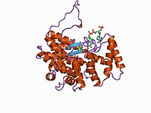
 cold-active citrate synthase | |||||||||
| Identifiers | |||||||||
| Symbol | Citrate_synt | ||||||||
| Pfam | PF00285 | ||||||||
| InterPro | IPR002020 | ||||||||
| PROSITE | PDOC00422 | ||||||||
| SCOP2 | 1csc / SCOPe / SUPFAM | ||||||||
| CDD | cd06101 | ||||||||
| |||||||||
In molecular biology, the citrate synthase family of proteins includes the enzymes citrate synthase EC 2.3.3.1, and the related enzymes 2-methylcitrate synthase EC 2.3.3.5 and ATP citrate lyase EC 2.3.3.8.
Citrate synthase is a member of a small family of enzymes that can directly form a carbon-carbon bond without the presence of metal ion cofactors. It catalyses the first reaction in the Krebs' cycle, namely the conversion of oxaloacetate and acetyl-coenzyme A into citrate and coenzyme A. This reaction is important for energy generation and for carbon assimilation. The reaction proceeds via a non-covalently bound citryl-coenzyme A intermediate in a 2-step process (aldol-Claisen condensation followed by the hydrolysis of citryl-CoA).
Citrate synthase enzymes are found in two distinct structural types: type I enzymes (found in eukaryotes, Gram-positive bacteria and archaea) form homodimers and have shorter sequences than type II enzymes, which are found in Gram-negative bacteria and are hexameric in structure. In both types, the monomer is composed of two domains: a large alpha-helical domain consisting of two structural repeats, where the second repeat is interrupted by a small alpha-helical domain. The cleft between these domains forms the active site, where both citrate and acetyl-coenzyme A bind. The enzyme undergoes a conformational change upon binding of the oxaloacetate ligand, whereby the active site cleft closes over in order to form the acetyl-CoA binding site. [1] The energy required for domain closure comes from the interaction of the enzyme with the substrate. Type II enzymes possess an extra N-terminal beta-sheet domain, and some type II enzymes are allosterically inhibited by NADH. [2]
2-methylcitrate synthase catalyses the conversion of oxaloacetate and propanoyl-CoA into (2R,3S)-2-hydroxybutane-1,2,3-tricarboxylate and coenzyme A. This enzyme is induced during bacterial growth on propionate, while type II hexameric citrate synthase is constitutive. [3]
ATP citrate lyase catalyses the Mg.ATP-dependent, CoA-dependent cleavage of citrate into oxaloacetate and acetyl-CoA, a key step in the reductive tricarboxylic acid pathway of CO2 assimilation used by a variety of autotrophic bacteria and archaea to fix carbon dioxide. [4] ATP citrate lyase is composed of two distinct subunits. In eukaryotes, ATP citrate lyase is a homotetramer of a single large polypeptide, and is used to produce cytosolic acetyl-CoA from mitochondrial produced citrate. [5]
References
- ^ Daidone I, Roccatano D, Hayward S (June 2004). "Investigating the accessibility of the closed domain conformation of citrate synthase using essential dynamics sampling". J. Mol. Biol. 339 (3): 515–25. doi: 10.1016/j.jmb.2004.04.007. PMID 15147839.
- ^ Francois JA, Starks CM, Sivanuntakorn S, Jiang H, Ransome AE, Nam JW, Constantine CZ, Kappock TJ (November 2006). "Structure of a NADH-insensitive hexameric citrate synthase that resists acid inactivation". Biochemistry. 45 (45): 13487–99. doi: 10.1021/bi061083k. PMID 17087502.
- ^ Gerike U, Hough DW, Russell NJ, Dyall-Smith ML, Danson MJ (April 1998). "Citrate synthase and 2-methylcitrate synthase: structural, functional and evolutionary relationships". Microbiology. 144 (4): 929–35. doi: 10.1099/00221287-144-4-929. PMID 9579066.
- ^ Kim W, Tabita FR (September 2006). "Both subunits of ATP-citrate lyase from Chlorobium tepidum contribute to catalytic activity". J. Bacteriol. 188 (18): 6544–52. doi: 10.1128/JB.00523-06. PMC 1595482. PMID 16952946.
- ^ Bauer DE, Hatzivassiliou G, Zhao F, Andreadis C, Thompson CB (September 2005). "ATP citrate lyase is an important component of cell growth and transformation". Oncogene. 24 (41): 6314–22. doi: 10.1038/sj.onc.1208773. PMID 16007201.
| Citrate_synt | |||||||||
|---|---|---|---|---|---|---|---|---|---|
 cold-active citrate synthase | |||||||||
| Identifiers | |||||||||
| Symbol | Citrate_synt | ||||||||
| Pfam | PF00285 | ||||||||
| InterPro | IPR002020 | ||||||||
| PROSITE | PDOC00422 | ||||||||
| SCOP2 | 1csc / SCOPe / SUPFAM | ||||||||
| CDD | cd06101 | ||||||||
| |||||||||
In molecular biology, the citrate synthase family of proteins includes the enzymes citrate synthase EC 2.3.3.1, and the related enzymes 2-methylcitrate synthase EC 2.3.3.5 and ATP citrate lyase EC 2.3.3.8.
Citrate synthase is a member of a small family of enzymes that can directly form a carbon-carbon bond without the presence of metal ion cofactors. It catalyses the first reaction in the Krebs' cycle, namely the conversion of oxaloacetate and acetyl-coenzyme A into citrate and coenzyme A. This reaction is important for energy generation and for carbon assimilation. The reaction proceeds via a non-covalently bound citryl-coenzyme A intermediate in a 2-step process (aldol-Claisen condensation followed by the hydrolysis of citryl-CoA).
Citrate synthase enzymes are found in two distinct structural types: type I enzymes (found in eukaryotes, Gram-positive bacteria and archaea) form homodimers and have shorter sequences than type II enzymes, which are found in Gram-negative bacteria and are hexameric in structure. In both types, the monomer is composed of two domains: a large alpha-helical domain consisting of two structural repeats, where the second repeat is interrupted by a small alpha-helical domain. The cleft between these domains forms the active site, where both citrate and acetyl-coenzyme A bind. The enzyme undergoes a conformational change upon binding of the oxaloacetate ligand, whereby the active site cleft closes over in order to form the acetyl-CoA binding site. [1] The energy required for domain closure comes from the interaction of the enzyme with the substrate. Type II enzymes possess an extra N-terminal beta-sheet domain, and some type II enzymes are allosterically inhibited by NADH. [2]
2-methylcitrate synthase catalyses the conversion of oxaloacetate and propanoyl-CoA into (2R,3S)-2-hydroxybutane-1,2,3-tricarboxylate and coenzyme A. This enzyme is induced during bacterial growth on propionate, while type II hexameric citrate synthase is constitutive. [3]
ATP citrate lyase catalyses the Mg.ATP-dependent, CoA-dependent cleavage of citrate into oxaloacetate and acetyl-CoA, a key step in the reductive tricarboxylic acid pathway of CO2 assimilation used by a variety of autotrophic bacteria and archaea to fix carbon dioxide. [4] ATP citrate lyase is composed of two distinct subunits. In eukaryotes, ATP citrate lyase is a homotetramer of a single large polypeptide, and is used to produce cytosolic acetyl-CoA from mitochondrial produced citrate. [5]
References
- ^ Daidone I, Roccatano D, Hayward S (June 2004). "Investigating the accessibility of the closed domain conformation of citrate synthase using essential dynamics sampling". J. Mol. Biol. 339 (3): 515–25. doi: 10.1016/j.jmb.2004.04.007. PMID 15147839.
- ^ Francois JA, Starks CM, Sivanuntakorn S, Jiang H, Ransome AE, Nam JW, Constantine CZ, Kappock TJ (November 2006). "Structure of a NADH-insensitive hexameric citrate synthase that resists acid inactivation". Biochemistry. 45 (45): 13487–99. doi: 10.1021/bi061083k. PMID 17087502.
- ^ Gerike U, Hough DW, Russell NJ, Dyall-Smith ML, Danson MJ (April 1998). "Citrate synthase and 2-methylcitrate synthase: structural, functional and evolutionary relationships". Microbiology. 144 (4): 929–35. doi: 10.1099/00221287-144-4-929. PMID 9579066.
- ^ Kim W, Tabita FR (September 2006). "Both subunits of ATP-citrate lyase from Chlorobium tepidum contribute to catalytic activity". J. Bacteriol. 188 (18): 6544–52. doi: 10.1128/JB.00523-06. PMC 1595482. PMID 16952946.
- ^ Bauer DE, Hatzivassiliou G, Zhao F, Andreadis C, Thompson CB (September 2005). "ATP citrate lyase is an important component of cell growth and transformation". Oncogene. 24 (41): 6314–22. doi: 10.1038/sj.onc.1208773. PMID 16007201.