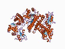
| CBL proto-oncogene N-terminal domain 1 (four-helix bundle) | |||||||||
|---|---|---|---|---|---|---|---|---|---|
 structure of the n-terminal domain of cbl in complex with its binding site in zap-70 | |||||||||
| Identifiers | |||||||||
| Symbol | Cbl_N | ||||||||
| Pfam | PF02262 | ||||||||
| InterPro | IPR003153 | ||||||||
| SCOP2 | 1b47 / SCOPe / SUPFAM | ||||||||
| |||||||||
| CBL proto-oncogene N-terminus, EF hand-like domain | |||||||||
|---|---|---|---|---|---|---|---|---|---|
 structure of the n-terminal domain of cbl in complex with its binding site in zap-70 | |||||||||
| Identifiers | |||||||||
| Symbol | Cbl_N2 | ||||||||
| Pfam | PF02761 | ||||||||
| InterPro | IPR014741 | ||||||||
| SCOP2 | 1b47 / SCOPe / SUPFAM | ||||||||
| |||||||||
| CBL proto-oncogene N-terminus, SH2-like domain | |||||||||
|---|---|---|---|---|---|---|---|---|---|
 structure of the n-terminal domain of cbl in complex with its binding site in zap-70 | |||||||||
| Identifiers | |||||||||
| Symbol | Cbl_N3 | ||||||||
| Pfam | PF02762 | ||||||||
| InterPro | IPR014742 | ||||||||
| SCOP2 | 1b47 / SCOPe / SUPFAM | ||||||||
| CDD | cd09920 | ||||||||
| |||||||||
In molecular biology, the Cbl TKB domain ( tyrosine kinase binding domain), also known as the phosphotyrosine binding (PTB) domain is a conserved region found at the N-terminus of Cbl adaptor proteins. This N-terminal region is composed of three evolutionarily conserved domains: an N-terminal four-helix bundle domain, an EF hand-like domain and a SH2-like domain, which together are known to bind to phosphorylated tyrosine residues. [1] [2]
References
- ^ Meng W, Sawasdikosol S, Burakoff SJ, Eck MJ (March 1999). "Structure of the amino-terminal domain of Cbl complexed to its binding site on ZAP-70 kinase". Nature. 398 (6722): 84–90. doi: 10.1038/18050. PMID 10078535.
- ^ Langenick J, Araki T, Yamada Y, Williams JG (November 2008). "A Dictyostelium homologue of the metazoan Cbl proteins regulates STAT signalling". Journal of Cell Science. 121 (Pt 21): 3524–30. doi: 10.1242/jcs.036798. PMID 18840649.
| CBL proto-oncogene N-terminal domain 1 (four-helix bundle) | |||||||||
|---|---|---|---|---|---|---|---|---|---|
 structure of the n-terminal domain of cbl in complex with its binding site in zap-70 | |||||||||
| Identifiers | |||||||||
| Symbol | Cbl_N | ||||||||
| Pfam | PF02262 | ||||||||
| InterPro | IPR003153 | ||||||||
| SCOP2 | 1b47 / SCOPe / SUPFAM | ||||||||
| |||||||||
| CBL proto-oncogene N-terminus, EF hand-like domain | |||||||||
|---|---|---|---|---|---|---|---|---|---|
 structure of the n-terminal domain of cbl in complex with its binding site in zap-70 | |||||||||
| Identifiers | |||||||||
| Symbol | Cbl_N2 | ||||||||
| Pfam | PF02761 | ||||||||
| InterPro | IPR014741 | ||||||||
| SCOP2 | 1b47 / SCOPe / SUPFAM | ||||||||
| |||||||||
| CBL proto-oncogene N-terminus, SH2-like domain | |||||||||
|---|---|---|---|---|---|---|---|---|---|
 structure of the n-terminal domain of cbl in complex with its binding site in zap-70 | |||||||||
| Identifiers | |||||||||
| Symbol | Cbl_N3 | ||||||||
| Pfam | PF02762 | ||||||||
| InterPro | IPR014742 | ||||||||
| SCOP2 | 1b47 / SCOPe / SUPFAM | ||||||||
| CDD | cd09920 | ||||||||
| |||||||||
In molecular biology, the Cbl TKB domain ( tyrosine kinase binding domain), also known as the phosphotyrosine binding (PTB) domain is a conserved region found at the N-terminus of Cbl adaptor proteins. This N-terminal region is composed of three evolutionarily conserved domains: an N-terminal four-helix bundle domain, an EF hand-like domain and a SH2-like domain, which together are known to bind to phosphorylated tyrosine residues. [1] [2]
References
- ^ Meng W, Sawasdikosol S, Burakoff SJ, Eck MJ (March 1999). "Structure of the amino-terminal domain of Cbl complexed to its binding site on ZAP-70 kinase". Nature. 398 (6722): 84–90. doi: 10.1038/18050. PMID 10078535.
- ^ Langenick J, Araki T, Yamada Y, Williams JG (November 2008). "A Dictyostelium homologue of the metazoan Cbl proteins regulates STAT signalling". Journal of Cell Science. 121 (Pt 21): 3524–30. doi: 10.1242/jcs.036798. PMID 18840649.