| Cytochrome c oxidase subunit Vb | |||||||||||
|---|---|---|---|---|---|---|---|---|---|---|---|
 Structure of the 13-subunit oxidized cytochrome c oxidase.
[1] | |||||||||||
| Identifiers | |||||||||||
| Symbol | COX5B | ||||||||||
| Pfam | PF01215 | ||||||||||
| InterPro | IPR002124 | ||||||||||
| PROSITE | PDOC00663 | ||||||||||
| SCOP2 | 1occ / SCOPe / SUPFAM | ||||||||||
| OPM superfamily | 4 | ||||||||||
| OPM protein | 1v55 | ||||||||||
| CDD | cd00924 | ||||||||||
| |||||||||||
Cytochrome c oxidase subunit 5B, mitochondrial is an enzyme in humans that is a subunit of the cytochrome c oxidase complex, also known as Complex IV, the last enzyme in the mitochondrial electron transport chain. [2] In humans, cytochrome c oxidase subunit 5B is encoded by the COX5B gene.
Structure
The enzyme weighs 14 kDa and is composed of 129 amino acids. [3] [4] The protein is a subunit of Complex IV, which consists of 13 mitochondrial- and nuclear-encoded subunits. [2] The sequence of subunit Vb is well conserved and includes three conserved cysteines that coordinate the zinc ion. [5] [6] Two of these cysteines are clustered in the C-terminal section of the subunit.
Gene
| COX5B | |||||||||||||||||||||||||||||||||||||||||||||||||||
|---|---|---|---|---|---|---|---|---|---|---|---|---|---|---|---|---|---|---|---|---|---|---|---|---|---|---|---|---|---|---|---|---|---|---|---|---|---|---|---|---|---|---|---|---|---|---|---|---|---|---|---|
| Identifiers | |||||||||||||||||||||||||||||||||||||||||||||||||||
| Aliases | COX5B, cytochrome c oxidase subunit Vb, COXVB, cytochrome c oxidase subunit 5B | ||||||||||||||||||||||||||||||||||||||||||||||||||
| External IDs | OMIM: 123866; MGI: 88475; HomoloGene: 37538; GeneCards: COX5B; OMA: COX5B - orthologs | ||||||||||||||||||||||||||||||||||||||||||||||||||
| |||||||||||||||||||||||||||||||||||||||||||||||||||
| |||||||||||||||||||||||||||||||||||||||||||||||||||
| |||||||||||||||||||||||||||||||||||||||||||||||||||
| |||||||||||||||||||||||||||||||||||||||||||||||||||
| |||||||||||||||||||||||||||||||||||||||||||||||||||
| Wikidata | |||||||||||||||||||||||||||||||||||||||||||||||||||
| |||||||||||||||||||||||||||||||||||||||||||||||||||
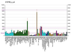
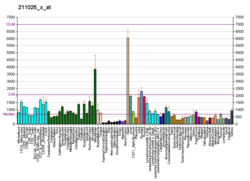
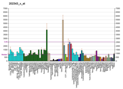
The COX5B gene, located on the q arm of chromosome 2 in position 11.2, is made up of 4 exons and is 2,137 base pairs in length. [2]
Function
Cytochrome c oxidase (COX) is the terminal enzyme of the mitochondrial respiratory chain. It is a multi-subunit enzyme complex that couples the transfer of electrons from cytochrome c to oxygen and contributes to a proton electrochemical gradient across the inner mitochondrial membrane to drive ATP synthesis via protonmotive force. The mitochondrially-encoded subunits perform the electron transfer of proton pumping activities. The functions of the nuclear-encoded subunits are unknown but they may play a role in the regulation and assembly of the complex. [2]
Summary reaction:
- 4 Fe2+-cytochrome c + 8 H+in + O2 → 4 Fe3+-cytochrome c + 2 H2O + 4 H+out [11]
Clinical significance
COX5A and COX5B are involved in the regulation of cancer cell metabolism by Bcl-2. [12]
The Trans-activator of transcription protein (Tat) of human immunodeficiency virus (HIV) inhibits cytochrome c oxidase (COX) activity in permeabilized mitochondria isolated from both mouse and human liver, heart, and brain samples. [13]
Interactions
COX5B has been shown to interact with Androgen receptor. [14]
References
- ^ Miki K, Sogabe S, Uno A, et al. (May 1994). "Application of an automatic molecular-replacement procedure to crystal structure analysis of cytochrome c2 from Rhodopseudomonas viridis" (PDF). Acta Crystallogr. D. 50 (Pt 3): 271–5. Bibcode: 1994AcCrD..50..271M. doi: 10.1107/S0907444993013952. PMID 15299438.
- ^ a b c d "Entrez Gene: COX5B cytochrome c oxidase subunit Vb".
- ^ Zong NC, Li H, Li H, Lam MP, Jimenez RC, Kim CS, Deng N, Kim AK, Choi JH, Zelaya I, Liem D, Meyer D, Odeberg J, Fang C, Lu HJ, Xu T, Weiss J, Duan H, Uhlen M, Yates JR, Apweiler R, Ge J, Hermjakob H, Ping P (Oct 2013). "Integration of cardiac proteome biology and medicine by a specialized knowledgebase". Circulation Research. 113 (9): 1043–53. doi: 10.1161/CIRCRESAHA.113.301151. PMC 4076475. PMID 23965338.
- ^ "Cytochrome c oxidase subunit 5B, mitochondrial". Cardiac Organellar Protein Atlas Knowledgebase (COPaKB). Archived from the original on 2018-07-19. Retrieved 2018-07-18.
- ^ Rizzuto R, Sandona D, Brini M, Capaldi RA, Bisson R (1991). "The most conserved nuclear-encoded polypeptide of cytochrome c oxidase is the putative zinc-binding subunit: primary structure of subunit V from the slime mold Dictyostelium discoideum". Biochim. Biophys. Acta. 1129 (1): 100–104. doi: 10.1016/0167-4781(91)90220-G. PMID 1661610.
- ^ Tsukihara T, Yamaguchi H, Aoyama H, Yamashita E, Tomizaki T, Shinzawa-Itoh K, Nakashima R, Yaono R, Yoshikawa S (1996). "The whole structure of the 13-subunit oxidized cytochrome c oxidase at 2.8 A". Science. 272 (5265): 1136–1144. Bibcode: 1996Sci...272.1136T. doi: 10.1126/science.272.5265.1136. PMID 8638158. S2CID 20860573.
- ^ a b c GRCh38: Ensembl release 89: ENSG00000135940 – Ensembl, May 2017
- ^ a b c GRCm38: Ensembl release 89: ENSMUSG00000061518 – Ensembl, May 2017
- ^ "Human PubMed Reference:". National Center for Biotechnology Information, U.S. National Library of Medicine.
- ^ "Mouse PubMed Reference:". National Center for Biotechnology Information, U.S. National Library of Medicine.
-
^ Pratt Donald Voet, Judith G. Voet, Charlotte W. (2013). "18". Fundamentals of biochemistry : life at the molecular level (4th ed.). Hoboken, NJ: Wiley. pp. 581–620.
ISBN
978-0-470-54784-7.
{{
cite book}}
: CS1 maint: multiple names: authors list ( link) - ^ Chen ZX, Pervaiz S (Mar 2010). "Involvement of cytochrome c oxidase subunits Va and Vb in the regulation of cancer cell metabolism by Bcl-2". Cell Death and Differentiation. 17 (3): 408–20. doi: 10.1038/cdd.2009.132. PMID 19834492.
- ^ Lecoeur H, Borgne-Sanchez A, Chaloin O, El-Khoury R, Brabant M, Langonné A, Porceddu M, Brière JJ, Buron N, Rebouillat D, Péchoux C, Deniaud A, Brenner C, Briand JP, Muller S, Rustin P, Jacotot E (2012). "HIV-1 Tat protein directly induces mitochondrial membrane permeabilization and inactivates cytochrome c oxidase". Cell Death & Disease. 3 (3): e282. doi: 10.1038/cddis.2012.21. PMC 3317353. PMID 22419111.
- ^ Beauchemin AM, Gottlieb B, Beitel LK, Elhaji YA, Pinsky L, Trifiro MA (2001). "Cytochrome c oxidase subunit Vb interacts with human androgen receptor: a potential mechanism for neurotoxicity in spinobulbar muscular atrophy". Brain Res. Bull. 56 (3–4): 285–97. doi: 10.1016/S0361-9230(01)00583-4. PMID 11719263. S2CID 24740136.
Further reading
- Lomax MI, Hsieh CL, Darras BT, Francke U (1991). "Structure of the human cytochrome c oxidase subunit Vb gene and chromosomal mapping of the coding gene and of seven pseudogenes". Genomics. 10 (1): 1–9. doi: 10.1016/0888-7543(91)90476-U. hdl: 2027.42/29338. PMID 1646156.
- Romero N, Marsac C, Fardeau M, Droste M, Schneyder B, Kadenbach B (1990). "Immunohistochemical demonstration of fibre type-specific isozymes of cytochrome c oxidase in human skeletal muscle". Histochemistry. 94 (2): 211–5. doi: 10.1007/BF02440190. PMID 2162812. S2CID 33365867.
- Zeviani M, Sakoda S, Sherbany AA, Nakase H, Rizzuto R, Samitt CE, DiMauro S, Schon EA (1988). "Sequence of cDNAs encoding subunit Vb of human and bovine cytochrome c oxidase". Gene. 65 (1): 1–11. doi: 10.1016/0378-1119(88)90411-8. PMID 2840351.
- Hughes GJ, Frutiger S, Paquet N, Pasquali C, Sanchez JC, Tissot JD, Bairoch A, Appel RD, Hochstrasser DF (1993). "Human liver protein map: update 1993". Electrophoresis. 14 (11): 1216–22. doi: 10.1002/elps.11501401181. PMID 8313870. S2CID 33424554.
- Bachman NJ, Yang TL, Dasen JS, Ernst RE, Lomax MI (1996). "Phylogenetic footprinting of the human cytochrome c oxidase subunit VB promoter". Arch. Biochem. Biophys. 333 (1): 152–62. doi: 10.1006/abbi.1996.0376. PMID 8806766.
- Lefai E, Vincent A, Boespflug-Tanguy O, Tanguy A, Alziari S (1997). "Quantitative decrease of human cytochrome c oxidase during development: evidences for a post-transcriptional regulation". Biochim. Biophys. Acta. 1318 (1–2): 191–201. doi: 10.1016/S0005-2728(96)00136-3. PMID 9030264.
- Wu H, Rao GN, Dai B, Singh P (2000). "Autocrine gastrins in colon cancer cells Up-regulate cytochrome c oxidase Vb and down-regulate efflux of cytochrome c and activation of caspase-3". J. Biol. Chem. 275 (42): 32491–8. doi: 10.1074/jbc.M002458200. PMID 10915781.
- Beauchemin AM, Gottlieb B, Beitel LK, Elhaji YA, Pinsky L, Trifiro MA (2002). "Cytochrome c oxidase subunit Vb interacts with human androgen receptor: a potential mechanism for neurotoxicity in spinobulbar muscular atrophy". Brain Res. Bull. 56 (3–4): 285–97. doi: 10.1016/S0361-9230(01)00583-4. PMID 11719263. S2CID 24740136.
- Ewing RM, Chu P, Elisma F, Li H, Taylor P, Climie S, McBroom-Cerajewski L, Robinson MD, O'Connor L, Li M, Taylor R, Dharsee M, Ho Y, Heilbut A, Moore L, Zhang S, Ornatsky O, Bukhman YV, Ethier M, Sheng Y, Vasilescu J, Abu-Farha M, Lambert JP, Duewel HS, Stewart II, Kuehl B, Hogue K, Colwill K, Gladwish K, Muskat B, Kinach R, Adams SL, Moran MF, Morin GB, Topaloglou T, Figeys D (2007). "Large-scale mapping of human protein-protein interactions by mass spectrometry". Mol. Syst. Biol. 3 (1): 89. doi: 10.1038/msb4100134. PMC 1847948. PMID 17353931.
External links
- Human COX5B genome location and COX5B gene details page in the UCSC Genome Browser.
- Mass spectrometry characterization of COX5B at COPaKB
- Cytochrome c oxidase subunit Vb in PROSITE
- Overview of all the structural information available in the PDB for UniProt: P10606 (Cytochrome c oxidase subunit 5B, mitochondrial) at the PDBe-KB.
This article incorporates text from the United States National Library of Medicine, which is in the public domain.
| Cytochrome c oxidase subunit Vb | |||||||||||
|---|---|---|---|---|---|---|---|---|---|---|---|
 Structure of the 13-subunit oxidized cytochrome c oxidase.
[1] | |||||||||||
| Identifiers | |||||||||||
| Symbol | COX5B | ||||||||||
| Pfam | PF01215 | ||||||||||
| InterPro | IPR002124 | ||||||||||
| PROSITE | PDOC00663 | ||||||||||
| SCOP2 | 1occ / SCOPe / SUPFAM | ||||||||||
| OPM superfamily | 4 | ||||||||||
| OPM protein | 1v55 | ||||||||||
| CDD | cd00924 | ||||||||||
| |||||||||||
Cytochrome c oxidase subunit 5B, mitochondrial is an enzyme in humans that is a subunit of the cytochrome c oxidase complex, also known as Complex IV, the last enzyme in the mitochondrial electron transport chain. [2] In humans, cytochrome c oxidase subunit 5B is encoded by the COX5B gene.
Structure
The enzyme weighs 14 kDa and is composed of 129 amino acids. [3] [4] The protein is a subunit of Complex IV, which consists of 13 mitochondrial- and nuclear-encoded subunits. [2] The sequence of subunit Vb is well conserved and includes three conserved cysteines that coordinate the zinc ion. [5] [6] Two of these cysteines are clustered in the C-terminal section of the subunit.
Gene
| COX5B | |||||||||||||||||||||||||||||||||||||||||||||||||||
|---|---|---|---|---|---|---|---|---|---|---|---|---|---|---|---|---|---|---|---|---|---|---|---|---|---|---|---|---|---|---|---|---|---|---|---|---|---|---|---|---|---|---|---|---|---|---|---|---|---|---|---|
| Identifiers | |||||||||||||||||||||||||||||||||||||||||||||||||||
| Aliases | COX5B, cytochrome c oxidase subunit Vb, COXVB, cytochrome c oxidase subunit 5B | ||||||||||||||||||||||||||||||||||||||||||||||||||
| External IDs | OMIM: 123866; MGI: 88475; HomoloGene: 37538; GeneCards: COX5B; OMA: COX5B - orthologs | ||||||||||||||||||||||||||||||||||||||||||||||||||
| |||||||||||||||||||||||||||||||||||||||||||||||||||
| |||||||||||||||||||||||||||||||||||||||||||||||||||
| |||||||||||||||||||||||||||||||||||||||||||||||||||
| |||||||||||||||||||||||||||||||||||||||||||||||||||
| |||||||||||||||||||||||||||||||||||||||||||||||||||
| Wikidata | |||||||||||||||||||||||||||||||||||||||||||||||||||
| |||||||||||||||||||||||||||||||||||||||||||||||||||
The COX5B gene, located on the q arm of chromosome 2 in position 11.2, is made up of 4 exons and is 2,137 base pairs in length. [2]
Function
Cytochrome c oxidase (COX) is the terminal enzyme of the mitochondrial respiratory chain. It is a multi-subunit enzyme complex that couples the transfer of electrons from cytochrome c to oxygen and contributes to a proton electrochemical gradient across the inner mitochondrial membrane to drive ATP synthesis via protonmotive force. The mitochondrially-encoded subunits perform the electron transfer of proton pumping activities. The functions of the nuclear-encoded subunits are unknown but they may play a role in the regulation and assembly of the complex. [2]
Summary reaction:
- 4 Fe2+-cytochrome c + 8 H+in + O2 → 4 Fe3+-cytochrome c + 2 H2O + 4 H+out [11]
Clinical significance
COX5A and COX5B are involved in the regulation of cancer cell metabolism by Bcl-2. [12]
The Trans-activator of transcription protein (Tat) of human immunodeficiency virus (HIV) inhibits cytochrome c oxidase (COX) activity in permeabilized mitochondria isolated from both mouse and human liver, heart, and brain samples. [13]
Interactions
COX5B has been shown to interact with Androgen receptor. [14]
References
- ^ Miki K, Sogabe S, Uno A, et al. (May 1994). "Application of an automatic molecular-replacement procedure to crystal structure analysis of cytochrome c2 from Rhodopseudomonas viridis" (PDF). Acta Crystallogr. D. 50 (Pt 3): 271–5. Bibcode: 1994AcCrD..50..271M. doi: 10.1107/S0907444993013952. PMID 15299438.
- ^ a b c d "Entrez Gene: COX5B cytochrome c oxidase subunit Vb".
- ^ Zong NC, Li H, Li H, Lam MP, Jimenez RC, Kim CS, Deng N, Kim AK, Choi JH, Zelaya I, Liem D, Meyer D, Odeberg J, Fang C, Lu HJ, Xu T, Weiss J, Duan H, Uhlen M, Yates JR, Apweiler R, Ge J, Hermjakob H, Ping P (Oct 2013). "Integration of cardiac proteome biology and medicine by a specialized knowledgebase". Circulation Research. 113 (9): 1043–53. doi: 10.1161/CIRCRESAHA.113.301151. PMC 4076475. PMID 23965338.
- ^ "Cytochrome c oxidase subunit 5B, mitochondrial". Cardiac Organellar Protein Atlas Knowledgebase (COPaKB). Archived from the original on 2018-07-19. Retrieved 2018-07-18.
- ^ Rizzuto R, Sandona D, Brini M, Capaldi RA, Bisson R (1991). "The most conserved nuclear-encoded polypeptide of cytochrome c oxidase is the putative zinc-binding subunit: primary structure of subunit V from the slime mold Dictyostelium discoideum". Biochim. Biophys. Acta. 1129 (1): 100–104. doi: 10.1016/0167-4781(91)90220-G. PMID 1661610.
- ^ Tsukihara T, Yamaguchi H, Aoyama H, Yamashita E, Tomizaki T, Shinzawa-Itoh K, Nakashima R, Yaono R, Yoshikawa S (1996). "The whole structure of the 13-subunit oxidized cytochrome c oxidase at 2.8 A". Science. 272 (5265): 1136–1144. Bibcode: 1996Sci...272.1136T. doi: 10.1126/science.272.5265.1136. PMID 8638158. S2CID 20860573.
- ^ a b c GRCh38: Ensembl release 89: ENSG00000135940 – Ensembl, May 2017
- ^ a b c GRCm38: Ensembl release 89: ENSMUSG00000061518 – Ensembl, May 2017
- ^ "Human PubMed Reference:". National Center for Biotechnology Information, U.S. National Library of Medicine.
- ^ "Mouse PubMed Reference:". National Center for Biotechnology Information, U.S. National Library of Medicine.
-
^ Pratt Donald Voet, Judith G. Voet, Charlotte W. (2013). "18". Fundamentals of biochemistry : life at the molecular level (4th ed.). Hoboken, NJ: Wiley. pp. 581–620.
ISBN
978-0-470-54784-7.
{{
cite book}}
: CS1 maint: multiple names: authors list ( link) - ^ Chen ZX, Pervaiz S (Mar 2010). "Involvement of cytochrome c oxidase subunits Va and Vb in the regulation of cancer cell metabolism by Bcl-2". Cell Death and Differentiation. 17 (3): 408–20. doi: 10.1038/cdd.2009.132. PMID 19834492.
- ^ Lecoeur H, Borgne-Sanchez A, Chaloin O, El-Khoury R, Brabant M, Langonné A, Porceddu M, Brière JJ, Buron N, Rebouillat D, Péchoux C, Deniaud A, Brenner C, Briand JP, Muller S, Rustin P, Jacotot E (2012). "HIV-1 Tat protein directly induces mitochondrial membrane permeabilization and inactivates cytochrome c oxidase". Cell Death & Disease. 3 (3): e282. doi: 10.1038/cddis.2012.21. PMC 3317353. PMID 22419111.
- ^ Beauchemin AM, Gottlieb B, Beitel LK, Elhaji YA, Pinsky L, Trifiro MA (2001). "Cytochrome c oxidase subunit Vb interacts with human androgen receptor: a potential mechanism for neurotoxicity in spinobulbar muscular atrophy". Brain Res. Bull. 56 (3–4): 285–97. doi: 10.1016/S0361-9230(01)00583-4. PMID 11719263. S2CID 24740136.
Further reading
- Lomax MI, Hsieh CL, Darras BT, Francke U (1991). "Structure of the human cytochrome c oxidase subunit Vb gene and chromosomal mapping of the coding gene and of seven pseudogenes". Genomics. 10 (1): 1–9. doi: 10.1016/0888-7543(91)90476-U. hdl: 2027.42/29338. PMID 1646156.
- Romero N, Marsac C, Fardeau M, Droste M, Schneyder B, Kadenbach B (1990). "Immunohistochemical demonstration of fibre type-specific isozymes of cytochrome c oxidase in human skeletal muscle". Histochemistry. 94 (2): 211–5. doi: 10.1007/BF02440190. PMID 2162812. S2CID 33365867.
- Zeviani M, Sakoda S, Sherbany AA, Nakase H, Rizzuto R, Samitt CE, DiMauro S, Schon EA (1988). "Sequence of cDNAs encoding subunit Vb of human and bovine cytochrome c oxidase". Gene. 65 (1): 1–11. doi: 10.1016/0378-1119(88)90411-8. PMID 2840351.
- Hughes GJ, Frutiger S, Paquet N, Pasquali C, Sanchez JC, Tissot JD, Bairoch A, Appel RD, Hochstrasser DF (1993). "Human liver protein map: update 1993". Electrophoresis. 14 (11): 1216–22. doi: 10.1002/elps.11501401181. PMID 8313870. S2CID 33424554.
- Bachman NJ, Yang TL, Dasen JS, Ernst RE, Lomax MI (1996). "Phylogenetic footprinting of the human cytochrome c oxidase subunit VB promoter". Arch. Biochem. Biophys. 333 (1): 152–62. doi: 10.1006/abbi.1996.0376. PMID 8806766.
- Lefai E, Vincent A, Boespflug-Tanguy O, Tanguy A, Alziari S (1997). "Quantitative decrease of human cytochrome c oxidase during development: evidences for a post-transcriptional regulation". Biochim. Biophys. Acta. 1318 (1–2): 191–201. doi: 10.1016/S0005-2728(96)00136-3. PMID 9030264.
- Wu H, Rao GN, Dai B, Singh P (2000). "Autocrine gastrins in colon cancer cells Up-regulate cytochrome c oxidase Vb and down-regulate efflux of cytochrome c and activation of caspase-3". J. Biol. Chem. 275 (42): 32491–8. doi: 10.1074/jbc.M002458200. PMID 10915781.
- Beauchemin AM, Gottlieb B, Beitel LK, Elhaji YA, Pinsky L, Trifiro MA (2002). "Cytochrome c oxidase subunit Vb interacts with human androgen receptor: a potential mechanism for neurotoxicity in spinobulbar muscular atrophy". Brain Res. Bull. 56 (3–4): 285–97. doi: 10.1016/S0361-9230(01)00583-4. PMID 11719263. S2CID 24740136.
- Ewing RM, Chu P, Elisma F, Li H, Taylor P, Climie S, McBroom-Cerajewski L, Robinson MD, O'Connor L, Li M, Taylor R, Dharsee M, Ho Y, Heilbut A, Moore L, Zhang S, Ornatsky O, Bukhman YV, Ethier M, Sheng Y, Vasilescu J, Abu-Farha M, Lambert JP, Duewel HS, Stewart II, Kuehl B, Hogue K, Colwill K, Gladwish K, Muskat B, Kinach R, Adams SL, Moran MF, Morin GB, Topaloglou T, Figeys D (2007). "Large-scale mapping of human protein-protein interactions by mass spectrometry". Mol. Syst. Biol. 3 (1): 89. doi: 10.1038/msb4100134. PMC 1847948. PMID 17353931.
External links
- Human COX5B genome location and COX5B gene details page in the UCSC Genome Browser.
- Mass spectrometry characterization of COX5B at COPaKB
- Cytochrome c oxidase subunit Vb in PROSITE
- Overview of all the structural information available in the PDB for UniProt: P10606 (Cytochrome c oxidase subunit 5B, mitochondrial) at the PDBe-KB.
This article incorporates text from the United States National Library of Medicine, which is in the public domain.


