| CHB HEX N-terminal domain | |||||||||
|---|---|---|---|---|---|---|---|---|---|
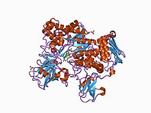
 beta-n-acetylhexosaminidase mutant e540d complexed with di-n acetyl-d-glucosamine (chitobiase) | |||||||||
| Identifiers | |||||||||
| Symbol | CHB_HEX | ||||||||
| Pfam | PF03173 | ||||||||
| Pfam clan | CL0203 | ||||||||
| InterPro | IPR004866 | ||||||||
| SCOP2 | 1c7s / SCOPe / SUPFAM | ||||||||
| |||||||||
In molecular biology, the CHB HEX N-terminal domain represents the N-terminal domain in chitobiases and beta-hexosaminidases. Chitobiases degrade chitin, which forms the exoskeleton in insects and crustaceans, and which is one of the most abundant polysaccharides on earth. [1] Beta-hexosaminidases are composed of either a HexA/HexB heterodimer or a HexB homodimer, and can hydrolyse diverse substrates, including GM(2)- gangliosides; mutations in this enzyme are associated with Tay–Sachs disease. [2] HexB is structurally similar to chitobiase, consisting of a beta sandwich structure; this structure is similar to that found in the cellulose-binding domain of cellulase from Cellulomonas fimi. [1] This domain may function as a carbohydrate binding module.
References
- ^ a b Tews I, Perrakis A, Oppenheim A, Dauter Z, Wilson KS, Vorgias CE (July 1996). "Bacterial chitobiase structure provides insight into catalytic mechanism and the basis of Tay–Sachs disease". Nat. Struct. Biol. 3 (7): 638–48. doi: 10.1038/nsb0796-638. PMID 8673609. S2CID 27327205.
- ^ Mark BL, Mahuran DJ, Cherney MM, Zhao D, Knapp S, James MN (April 2003). "Crystal structure of human beta-hexosaminidase B: understanding the molecular basis of Sandhoff and Tay–Sachs disease". J. Mol. Biol. 327 (5): 1093–109. doi: 10.1016/S0022-2836(03)00216-X. PMC 2910754. PMID 12662933.
| CHB HEX N-terminal domain | |||||||||
|---|---|---|---|---|---|---|---|---|---|
 beta-n-acetylhexosaminidase mutant e540d complexed with di-n acetyl-d-glucosamine (chitobiase) | |||||||||
| Identifiers | |||||||||
| Symbol | CHB_HEX | ||||||||
| Pfam | PF03173 | ||||||||
| Pfam clan | CL0203 | ||||||||
| InterPro | IPR004866 | ||||||||
| SCOP2 | 1c7s / SCOPe / SUPFAM | ||||||||
| |||||||||
In molecular biology, the CHB HEX N-terminal domain represents the N-terminal domain in chitobiases and beta-hexosaminidases. Chitobiases degrade chitin, which forms the exoskeleton in insects and crustaceans, and which is one of the most abundant polysaccharides on earth. [1] Beta-hexosaminidases are composed of either a HexA/HexB heterodimer or a HexB homodimer, and can hydrolyse diverse substrates, including GM(2)- gangliosides; mutations in this enzyme are associated with Tay–Sachs disease. [2] HexB is structurally similar to chitobiase, consisting of a beta sandwich structure; this structure is similar to that found in the cellulose-binding domain of cellulase from Cellulomonas fimi. [1] This domain may function as a carbohydrate binding module.
References
- ^ a b Tews I, Perrakis A, Oppenheim A, Dauter Z, Wilson KS, Vorgias CE (July 1996). "Bacterial chitobiase structure provides insight into catalytic mechanism and the basis of Tay–Sachs disease". Nat. Struct. Biol. 3 (7): 638–48. doi: 10.1038/nsb0796-638. PMID 8673609. S2CID 27327205.
- ^ Mark BL, Mahuran DJ, Cherney MM, Zhao D, Knapp S, James MN (April 2003). "Crystal structure of human beta-hexosaminidase B: understanding the molecular basis of Sandhoff and Tay–Sachs disease". J. Mol. Biol. 327 (5): 1093–109. doi: 10.1016/S0022-2836(03)00216-X. PMC 2910754. PMID 12662933.